Fig. 3.
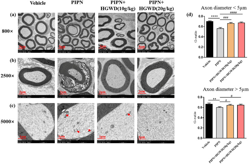
HGWD attenuated paclitaxel-induced histological changes in sciatic nerves. a Cross view of ultrastructure of rat sciatic nerve. Scale bar, 5 μm. b Paclitaxel-induced sciatic nerve damage includes axonal degeneration and myelin damage. Scale bar, 2 μm. c Representative transmission electron microscopy images show that paclitaxel caused swollen and vacuolated mitochondria (red arrow). Scale bar, 1 μm. d G-ratio (the ratio of inner axon circumference to outer myelin circumference) measurement in the sciatic nerves. Each value represents mean ± SEM of at least 32 sciatic nerves. **P < 0.01, ****P < 0.0001, vs. vehicle group; #P < 0.05, ###P < 0.001, ####P < 0.0001, vs. PIPN group

