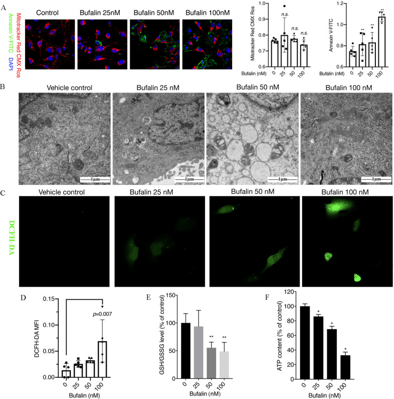
Fig. 1.
Bufalin induces cell apoptosis and oxidative stress. A U251 cells were treated with 25, 50 or 100 nM bufalin for 12 h, and cell apoptosis was observed by confocal microscopy after MitoTracker Red CMX ROS, Annexin V-FITC and Hoechst 33,342 co-staining (n = 3). The Y axis in MitoTracker Red CMX Ros and Annexin V-FITC plot represents the intensity of red and green fluorescence. B Ultrastructure of U251 cells as detected with TEM. C The intracellular ROS content was observed by laser confocal microscopy after DCFH-DA staining (n = 3). D The ROS levels in U251 cells. E The ratio of GSH/GSSH in each group of cells, **p < 0.01 compared with the DMSO control. F The intracellular ATP content in U251 cells after treatment with different concentrations of bufalin (n = 3). P values were determined using one-way ANOVA. The relative expression value for each sample is shown along with the mean ± SD for each group. *p < 0.05, **p < 0.01 compared with DMSO control

