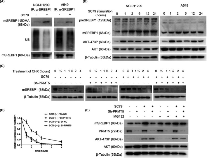
FIGURE 2.

Activation of protein kinase B (AKT) increases the protein arginine methyltransferase 5 (PRMT5)‐mediated protein stabilization of mature sterol regulatory element‐binding protein 1 (mSREBP1). A, After cells were cultured in medium without FBS for 24 h, SC79 (4 µg/mL; Selleck) was added (DMSO as control), and the cells were harvested at 24 h. Lysates of NCI‐H1299 or A549 cells were immunoprecipitated (IP) with anti‐SREBP1 Ab, and mSREBP1 expression levels were then normalized. Indicated Abs were used to analyze post‐translational modification of mSREBP1. UB, ubiquitin modification. B, After cells were cultured in medium without FBS for 24 h, SC79 (4 µg/mL; Selleck) was added (DMSO as control), and the cells were harvested at 0, 1, 2, 6, 12, and 24 h. Lysates of NCI‐H1299 or A549 cells were immunoblotted with Abs against SREBP1, Ser473‐phosphorylated AKT (AKT‐473P), or AKT. β‐Tubulin was used as a loading control. C, NCI‐H1299 cells with or without PRMT5 stable deletion were cultured in medium without FBS for 24 h. SC79 (4 µg/mL) was then added (DMSO as control) for 24 h. Subsequently, cycloheximide (CHX, 100 µg/mL; Sigma‐Aldrich) was added, and the cells were harvested at 0, 0.5, 1, 1.5, 2, and 4 h. Cell lysates were immunoblotted with an anti‐SREBP1 Ab. β‐Tubulin was used as a loading control. D, NCI‐H1299 cells with or without PRMT5 stable deletion were cultured in medium without FBS for 24 hours. SC79 (4 μg/ml) was then added (DMSO as control) for 24 h. Subsequently, CHX (100 μg/ml) was added, and the cells were harvested at 0, ½, 1, 1½, 2, and 4 h. The mSREBP1 degradation rate in the four NCI‐H1299 cell groups was then determined. The mSREBP1 expression level was normalized to the β‐Tubulin level before being normalized to the time = 0 control, and the degradation rates are presented as a percentage. This experiment was repeated for three times, and the mean and SD of degradation rates were shown. E, NCI‐H1299 cells with or without PRMT5 stable deletion were cultured in medium without FBS for 24 h. SC79 (4 µg/mL) was then added (DMSO as control) for 24 h. MG132 (100 µmol/L; Sigma‐Aldrich) was added, and cells were harvested at 2 h. Cell lysates were immunoblotted with Abs against SREBP1, PRMT5, AKT‐473P, or AKT. β‐Tubulin was used as a loading control
