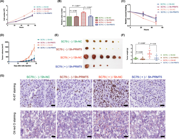
FIGURE 7.

Protein arginine methyltransferase 5 (PRMT5) knockdown reverses protein kinase B (AKT) activation‐induced growth of lung adenocarcinoma cells. A, NCI‐H1299 cells with or without PRMT5 stable deletion were cultured in medium without FBS for 24 h. SC79 (4 µg/mL) was then added (DMSO as control; Sh‐NC), and the number of cells was determined at 0, 24, 48, and 72 h. B, NCI‐H1299 cells with or without PRMT5 stable deletion were cultured in medium without FBS for 24 h. SC79 (4 µg/mL) was then added (DMSO as control), and cell colony formation assays were carried out at 14 days. The relative number of colonies was calculated, and the number of the control group was designated as 1. C, NCI‐H1299 cells with or without PRMT5 stable deletion were cultured in medium without FBS for 24 h. SC79 (4 µg/mL) was then added (DMSO as control), and cell wound‐healing assays were carried out at 0, 24, 48 h. Relative wound healing was normalized by the 0 h point of each group, which was designated 1. D‐G, Nude mice were injected with NCI‐H1299 cells with or without PRMT5 stable deletion and intraperitoneally injected with or without SC79 (100 mg/kg). D, Growth curve of in vivo tumor volume. E, Image of tumors isolated from nude mice. F, Weight of tumors when mice were killed. G, Representative images of Ki‐67 and oil red O staining of tumor samples. Scale bar = 50 μm
