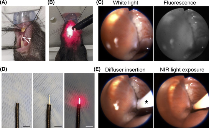
FIGURE 1.

Endoscopic procedures. A, MOC2‐luc cells were inoculated into the submucosal layer of the right buccal mucosa (arrowhead). B, An endoscope was inserted into the oral cavity. C, White light image and fluorescence image for IR700 are simultaneously displayed. D, A cylindrical light diffuser was inserted through the instrument channel of an endoscope. The diffuser was controlled by the endoscope. Left: tip of the endoscope, middle: inserted cylindrical diffuser, right: light exposure. Scale bars, 10 mm. E, In vivo NIR‐PIT. The light diffuser (*) was inserted (left) and the right buccal region was exposed to NIR light (right)
