
FIGURE 2.
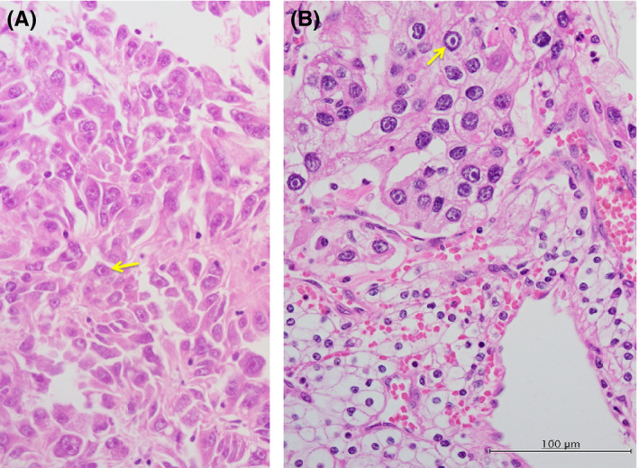
Prominent pale eosinophilic cytoplasmic inclusions within the tumor cells. Some neoplastic cells contained cytoplasmic eosinophilic inclusions. The tumor cells are pleomorphic appearance with cytoplasmic eosinophilic inclusions (yellow arrows) in case 2 (A) or enlarged, heavily stained (eosinophilic) nucleoli surrounded by a noticeable unstained space or halo within normally stained nuclei (yellow arrows) in case 8 (B)