FIGURE 1.

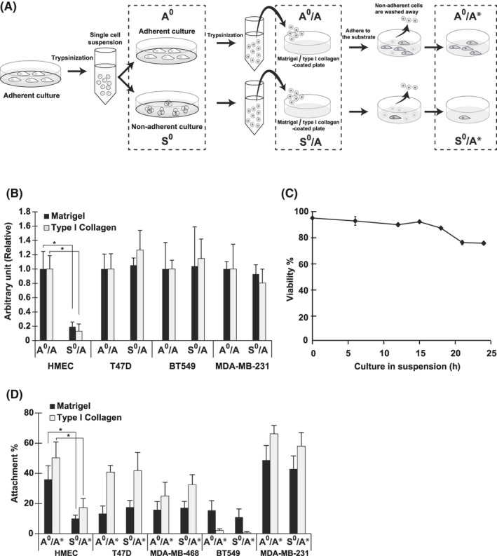
The colony forming ability (CFA) and reattachment ability (RA) of malignant and non‐malignant mammary epithelial cells under adherent and non‐adherent conditions. A, Schematic representation of the experimental procedure used in assays for attachment‐related properties of cells. Cells categorized into groups, adherent (A0) and non‐adherent (S0), and incubated for 24 h under A0 and S0 conditions, respectively, as described in Materials and Methods (cell lines and culture conditions), were used for western blotting. For colony formation and attachment assays, cells (A0 and S0) were dissociated and seeded onto plates coated with Matrigel or type I collagen (A0/A and S0/A). After 14 d, colony formation was quantified. The cell attachment assay was performed with cells (A0 and S0) dissociated and stained with the dye, as described in Materials and Methods. The cells were then replated and incubated for 30 min at A0/A and S0/A. After removal of unattached cells, the attached cells were quantified (A0/A* and S0/A*). Pull‐down assay was conducted following the steps for A0 and S0 or A0/A* and S0/A*. Representative images of cells (HMECs) cultured under each condition are shown in Figure S1A. Details for each assay are described in Materials and Methods. B, CFA of HMECs and cells from breast cancer cell lines (Table S1). Ratio to the control, the value from cells on Matrigel‐coated plates (A0/A; Matrigel) is shown. C, Viability of HMECs in suspension. HMECs were cultured under non‐adherent conditions (S0), and cell viability was determined by trypan blue exclusion at the time indicated. D, RAs of HMECs and cells from breast cancer cell lines assessed by cell attachment assays. Ratio of attached cells to the initial input is shown as attachment percentage. Values represent mean ± SD from at least 3 independent experiments. *P < .01
