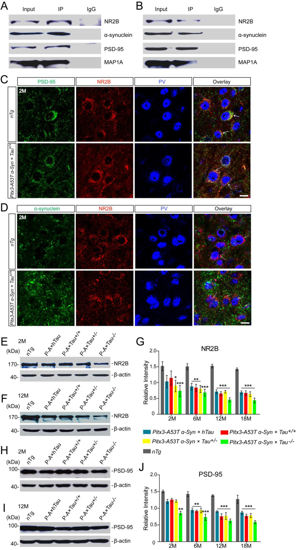
Figure. 6.

A53T α-syn mediated MAP1A impairment through the NR2B/PSD-95 pathway. (A,B) Representative immunoblotting showed the presence of α-syn, NR2B, PSD-95 and MAP1A positive bands in NR2B-immunoprecipitated protein fractions from the midbrain homogenates of 2 month-old nTg and Pitx3-A53T α-Syn × Tau+/+ mice, respectively. Mouse or rabbit IgG were used as negative controls. (C,D) Costaining images in the SNR of 2-month-old nTg and Pitx3-A53T α-Syn × Tau+/+ mice showed the apparent co-localization of PSD-95 and NR2B (C), α-syn and NR2B (D). Arrows pointed to the costainings. Scale bar: 10 μm. (E,F) Western blot showed the expression levels of NR2B in the midbrains of 2- and 12-month-old mice, respectively. (G) Bar graph illustrated the representative levels of NR2B. (H,I) Western blot showed the expression levels of PSD-95 in the midbrain homogenates of 2- and 12-month-old mice, respectively. (J) Bar graph showed the representative levels of PSD-95. Values are mean ± SEM. *P < 0.05, **P < 0.01, ***P < 0.001 (Triple transgenic versus age-matched nTg). n = 5 per genotype per time point.
