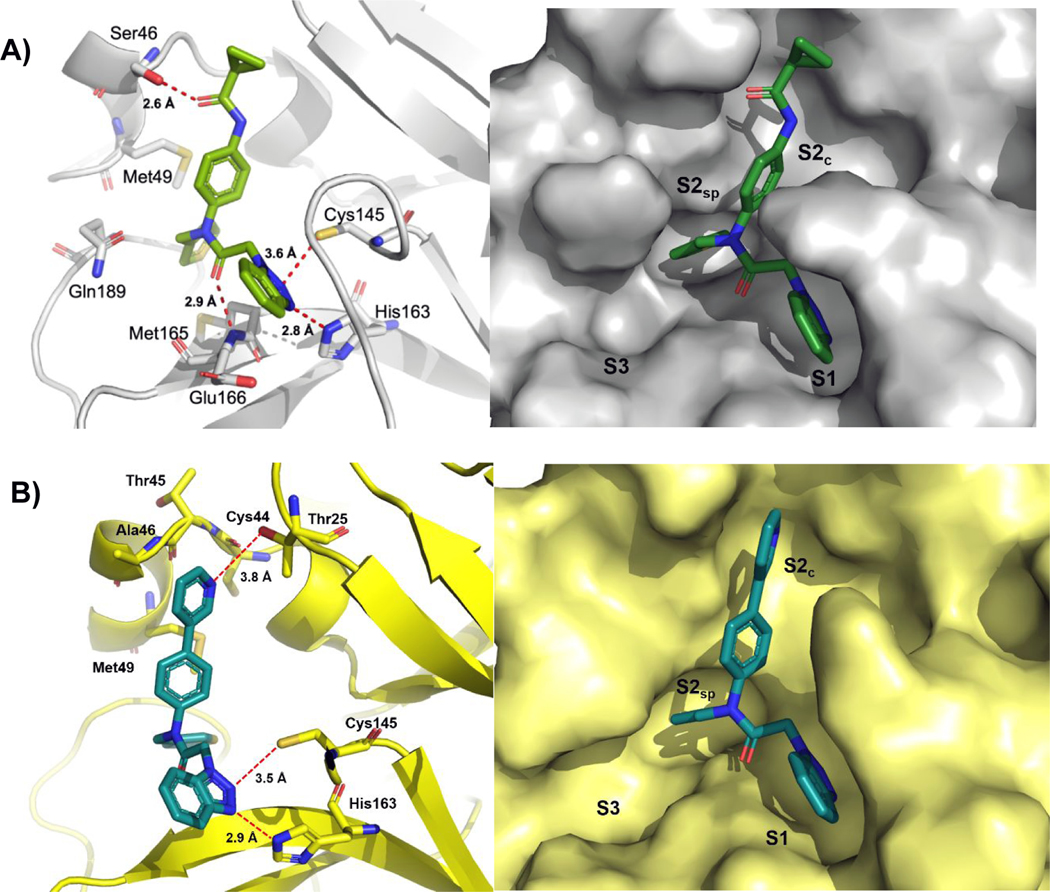
Figure 4.
A) X-ray co-crystal structure of 1 in complex with SC2 3CLpro (PDB: 7LME) and B) 8 in complex with SC1 3CLpro (PDB: 7LMH). Left panel: key residues are highlighted as sticks and interatomic distances depicted as dashes; right panel: solvent accessible surface with occupied and neighboring pockets labeled. Binding orientation of each inhibitor is the same in the active site of each monomer of the dimer in the asymmetric unit, only one active site is shown for clarity.

