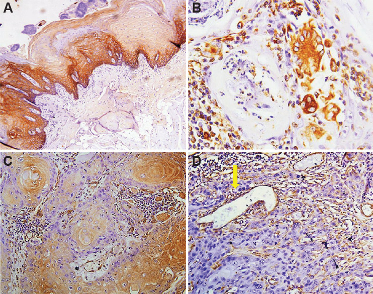
Fig. 2.

(A) Positive control for E-cadherin: expression in the adjacent normal mucosal epithelium (×100). (B) Vimentin positivity in the stromal cells, few lymphocytes and histiocytic collections acting as an internal control (×200). (C) E-cadherin expression seen at the centre of tumour islands in the most differentiated cells with loss in the peripheral cells and tumour buds (×100). (D) Vimentin positivity seen only in the stromal cells and vessels (arrow) and not in the tumour cells. This positivity acted as an internal control (×100).
