Fig. 3.

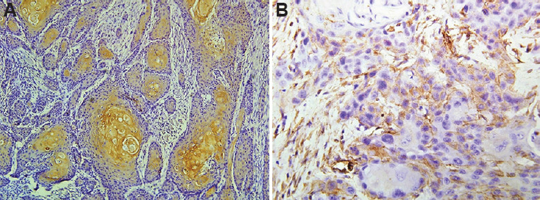
(A) Brown granular membranous E-cadherin positivity in centre of tumour islands (×200). (B) Vimentin positivity in cytoplasm of tumour cells at the infiltrating edge of the tumour (×200).

(A) Brown granular membranous E-cadherin positivity in centre of tumour islands (×200). (B) Vimentin positivity in cytoplasm of tumour cells at the infiltrating edge of the tumour (×200).