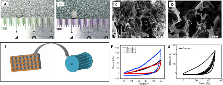
Figure 1.
Characterization of the collagen scaffold with axially-aligned luminal conduits.
(A, B) The collagen scaffold with a 3-mm diameter and 3-mm thickness, containing axially-aligned luminal conduits. (C, D) Scanning electron microscopy image showing the porous morphology of the collagen scaffold. Scale bars: 100 μm in C, 20 μm in D. (E) Schematic diagram of the mold for preparing the scaffold. (F, G) The compressive capacity of the collagen scaffold.

