Figure 4.
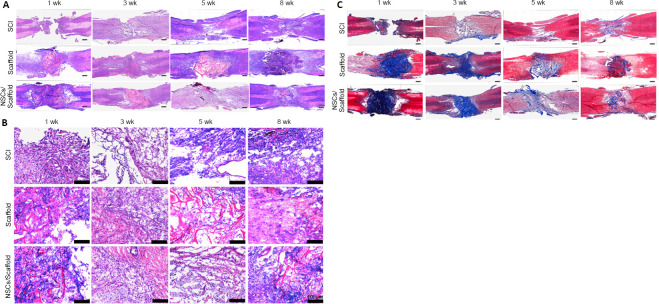
Effect of transplantation of collagen scaffold loaded with NSCs on the histopathology of the spinal cord in the SCI rat model.
(A, B) Hematoxylin and eosin staining of spinal cord tissue. The cavity at the lesion site in the collagen scaffold and the NSCs/collagen scaffold groups were significantly smaller than those in the SCI group. B is the enlarged view of the region in the dotted rectangle in A. (C) Masson staining of horizontal sections of spinal cords illustrating collagen deposits (blue). The collagen scaffolds in the collagen scaffold and the NSCs/collagen scaffold groups degraded over time. Scale bars: 500 μm in A, C; 100 μm in B. NSCs: Neural stem cells; SCI: spinal cord injury.

