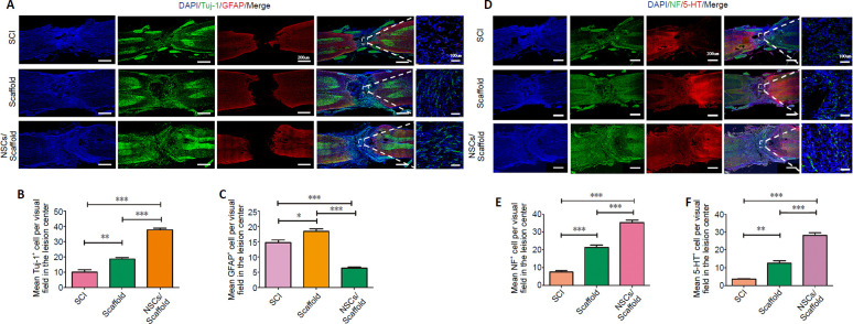
Figure 5.
NSCs/collagen scaffold treatment promotes nerve regeneration in SCI model rats.
(A) Immunofluorescence staining for Tuj-1 (Alexa Fluor 488, green) and GFAP (Alexa Fluor 568, red). The number of GFAP-positive astrocytes in the peri-lesional area in the SCI and collagen scaffold groups was higher than that in the NSCs/collagen scaffold group. Tuj-1-positive cells were more hypertrophic in the NSCs/collagen scaffold group compared with the SCI and collagen scaffold groups. (B, C, E, F) Quantification of Tuj-1-, GFAP-, NF- and 5-HT-positive cells in 200× field in SCI, collagen scaffold and NSCs/collagen scaffold groups. (D) Immunofluorescence staining for NF (Alexa Fluor 488, green) and 5-HT (Alexa Fluor 568, red) at the lesion site. The number of NF-positive and 5-HT-positive axons in the peri-lesional area in the NSCs/collagen scaffold group was higher than that in the other two groups. Data are presented as mean ± SD (n = 16). *P < 0.05, **P < 0.01, ***P < 0.001 (one-way analysis of variance followed by Tukey’s post hoc test). 5-HT: 5-Hydroxytryptamine; DAPI: 4',6-diamidino-2-phenylindole; GFAP: glial fibrillary acidic protein; NF: neurofilament; NSCs: neural stem cells; SCI: spinal cord injury; Tuj-1: βIII-tubulin.

