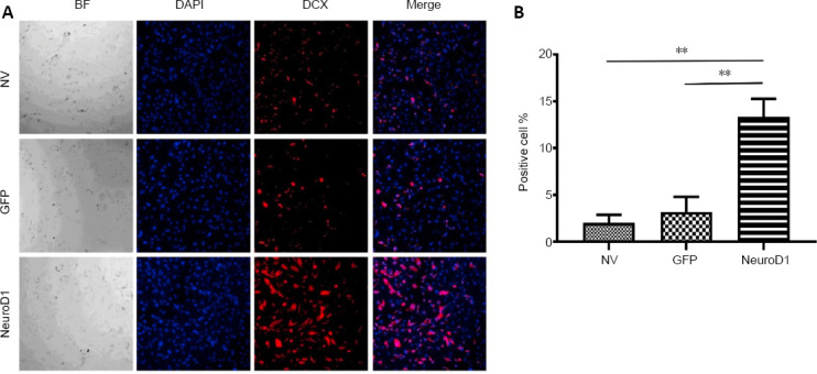
Figure 2.

NEUROD1-induced reactive astrocytes at 7 days after infection (immunofluorescence staining).
(A) NEUROD1 overexpression led to significantly increased DCX immunopositivity (red). (B) Percentage of DCX positive cells. Scale bar: 10 µm. Data are expressed as the mean ± SD. The experiment was repeated three times. **P < 0.01 (unpaired Student’s t-test). BF: Bright field; DAPI: 4′,6-diamidino-2-phenylindole; DCX: doublecortin; GFP: green fluorescent protein; NEUROD1: neurogenic differentiation factor 1; NV: non-virus group.
