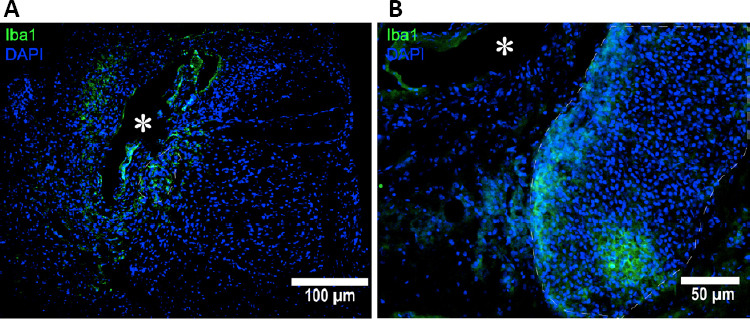
Figure 3.

Histological confirmation of premortem cyst formation using Iba1, microglia antibody marker (green) co-stained with DAPI (blue) for nuclei.
(A) Iba1-positive cells surrounding the cyst. (B) Cell aggregation adjacent to the cyst and it includes many Iba1-positive cells. The asterisk (*) indicates syrinx center. DAPI: 4′,6-Diamidino-2-phenylindole; Iba1: ionized calcium-binding adapter molecule 1.
