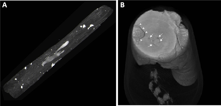
Figure 5.

Three-dimensional reconstruction of micro-computed tomography slices.
it shows a radio-opaque filled intraspinal cyst reconstructed within the spinal cord (A) and spinal cord segmented with a large cyst (B) marked with arrowheads and small defects within the spinal cord parenchyma.
