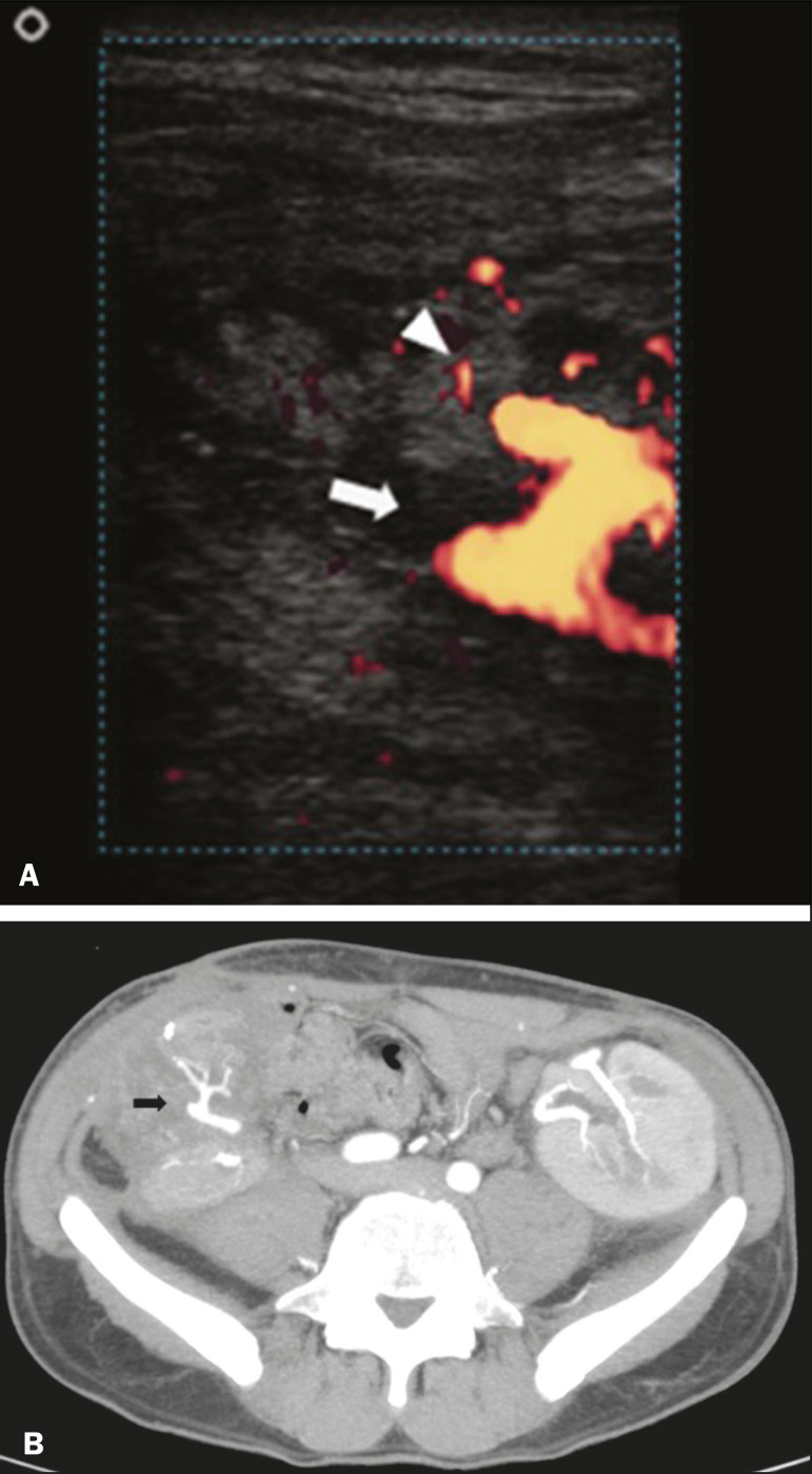
Figure 10.
A: Cross-sectional image obtained by ultrasound in power Doppler mode, showing abrupt interruption of flow in the distal segment of the superior mesenteric artery (arrow) of the pancreatic graft, with adjacent collateral circulation (arrowhead), a finding consistent with arterial thrombosis. B: Correlation with contrast-enhanced CT in the same patient, with maximum intensity projection in the arterial phase, identifying the same abrupt interruption and a filling defect in the distal segment of the superior mesenteric artery of the pancreatic graft (arrow), confirming the arterial thrombosis suggested by the ultrasound finding.

