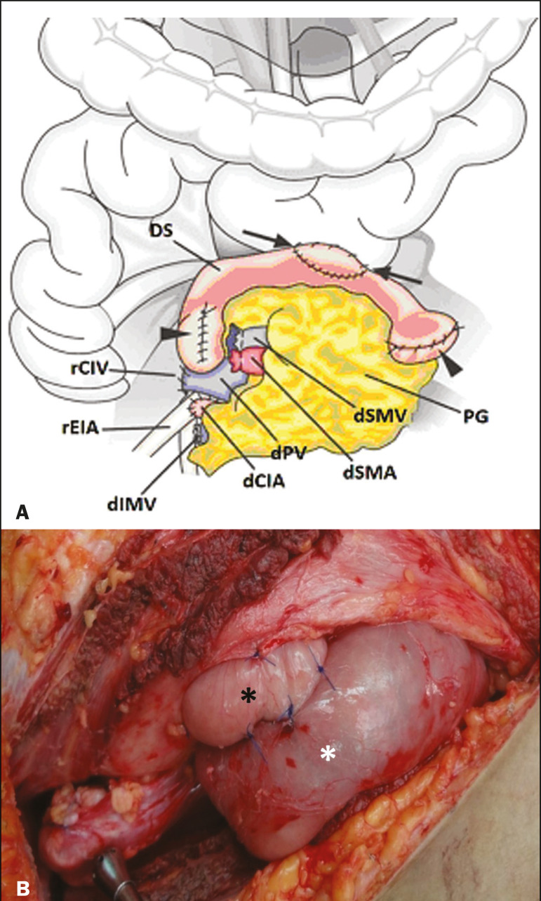
Figure 4.
A: Schematic drawing illustrating the intraoperative appearance of the pancreatic graft (PG) positioned in the pelvis via side-to-side duodenojejunal anastomosis (arrows) linking the donor duodenal segment (DS) to the jejunum of the recipient. The donor duodenum is closed at both ends using sutures or staples (arrowheads). The native pancreas of the recipient remains in the upper abdomen. B: Intraoperative aspect of SPKT with enteric drainage of exocrine pancreatic secretions, by anastomosis between the donor duodenal stump (black asterisk) and the small intestine of the recipient (white asterisk), approximately 25 cm from the ileocecal valve. dIMV, inferior mesenteric vein; dSMV, superior mesenteric vein.

