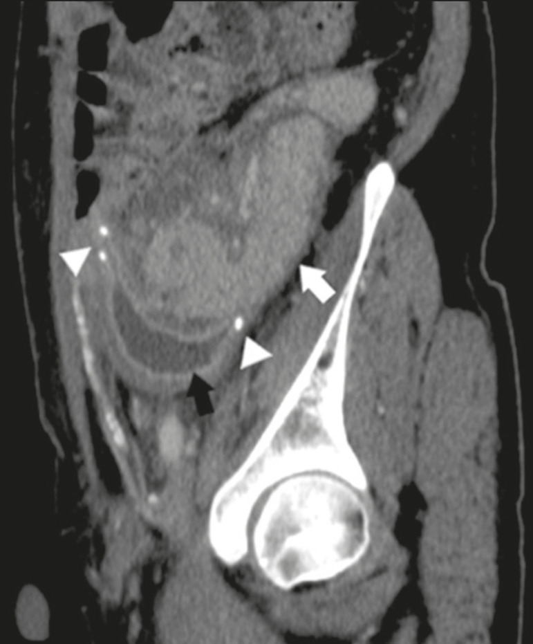
Figure 7.
Sagittal reconstruction of a contrast-enhanced CT scan of a pancreatic graft with enteric drainage in the right iliac fossa (white arrow), showing the surgical staples (arrowheads) along the duodenal stump (black arrow), which is partially distended and may simulate a fluid collection. Surgical staples facilitate this differentiation, as well as helping locate the pancreatic graft.

