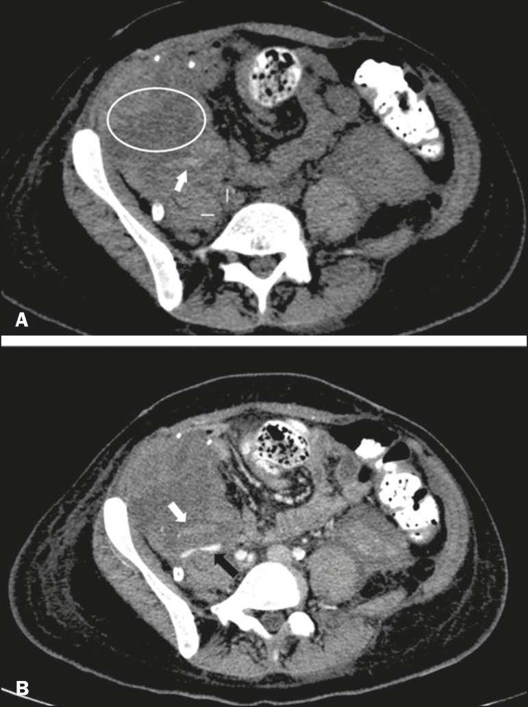
Figure 9.
A: Unenhanced abdominal CT scan in patient who underwent SPKT, showing a linear image of a spontaneously hyperattenuating mass (arrow) suggestive of thrombosis. In addition, the pancreatic graft is heterogeneous, with hypoattenuating areas (ellipse). B: Contrast-enhanced abdominal CT scan, in the venous phase, of the same patient, showing arterial opacification (black arrow) and the absence of venous opacification (white arrow), confirming the venous thrombosis seen in the previous phases.

