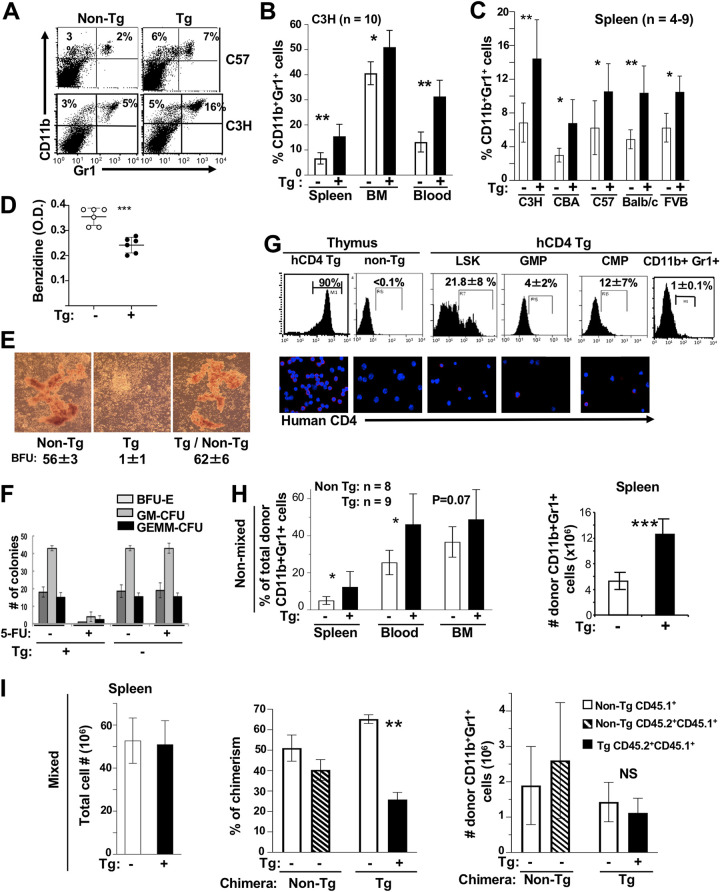
FIG 6.
Accumulation of CD11b+ Gr1+ cells in CD4C/Nef Tg mice. (A to C) Representative FACS profiles (A) and quantification (B and C) of CD11b+ Gr1+ cells in indicated organs of Tg and non-Tg mice of different backgrounds. Tg mice were bred for at least six generations as heterozygous on each mouse background before being studied. C57, C57BL/6. (D) Evaluation of benzidine-positive cells in BM of Tg and non-Tg mice. Total BM cells (1 × 10⁶) were suspended and stained with benzidine for 5 min and lysed, and the optical density at 420 nm (OD420) was measured. ***, P ≤ 0.001. (E) BM cells from control and CD4C/HIVMutA Tg mice or mixed mice (Tg/non-Tg) (1:1) were incubated with hydrocortisone (10−6 M) in methylcellulose and BFU-E colonies counted. (F) Impaired BM recovery after 5-FU treatment. Control non-Tg and CD4C/HIVMutA Tg mice were treated with 5-FU (100 mg/kg body weight) and sacrificed 10 days later. BM cells were cultured in methylcellulose and the number of colonies counted. (G) Expression of reporter human cell surface CD4 (hCD4) in myeloid progenitors and Gr1+ splenocytes of CD4/hCD4 Tg mice (n = 4). Expression in Tg and non-Tg thymus served as a positive and negative control, respectively. Myeloid progenitors and controls were labeled as shown in Fig. 4A and analyzed by FACS (upper) or by microscopic immunofluorescence (lower). (H and I) Non-Tg CD45.2 mice were lethally irradiated and transplanted, as described for Fig. 5I, with FL cells from CD45.1+ CD45.2+ Tg or non-Tg E14.5 embryos (nonmixed) (H) or with mixed (1:1) CD45.1+ CD45.2+ Tg/CD45.1+ non-Tg or control CD45.1+ CD45.2+ non-Tg/CD45.1+ non-Tg FL (I) cells. Recipient mice were analyzed 6 months posttransplantation. Results are from 3 independent experiments. *, P ≤ 0.05; **, P ≤ 0.01; ***, P ≤ 0.001.

