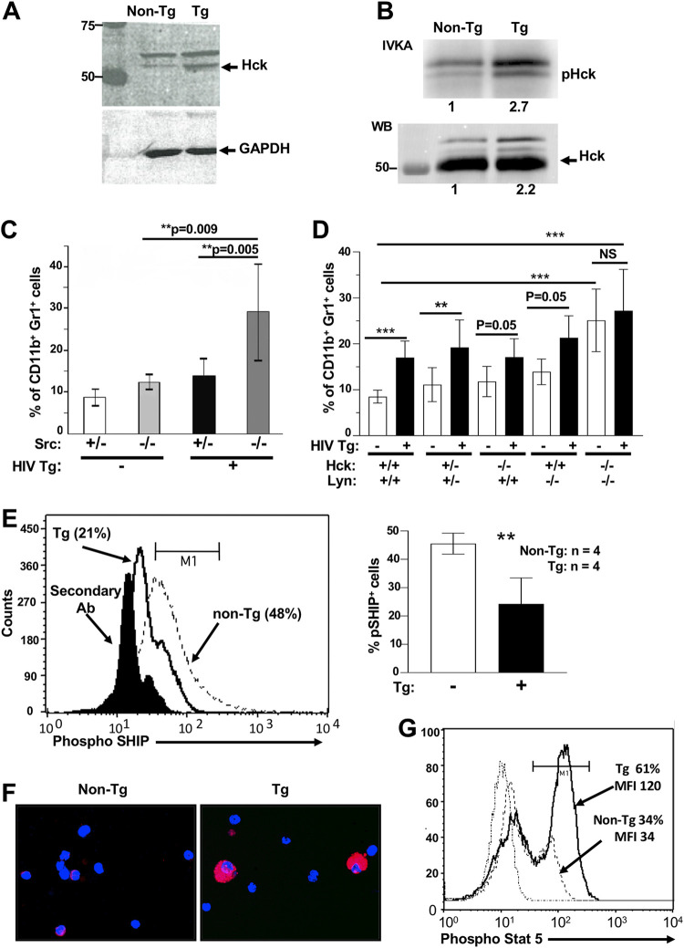
FIG 8.
Role of Src-related kinases in accumulation of CD11b+ Gr1+ myeloid cells of HIV Tg mice. (A and B) Lysates of spleen cells from CD11c/Nef Tg mice were analyzed by Western blotting (WB) with anti-Hck (A) or immunoprecipitated with anti-Hck Ab, and these immunoprecipitates were subjected to IVKA (B). Numbers below the graph refer to relative intensity. (C and D) Evaluation of percentage of CD11b+ Gr1+ cell subset in spleen of CD4C/Nef and CD4C/HIVMutA Tg mice bred on a wild-type (+/+), heterozygote (+/−), or homozygote (−/−) Src (n = 4 to 8/group) (C) or Hck/Lyn (n = 3 to 12/group) (D) gene-deficient background. (E) Evaluation of p-SHIPY1060 levels in unplated, freshly isolated purified peritoneal macrophages of Tg and non-Tg mice. Cells were labeled intracellularly with anti-p-SHIPY1060 Ab and Alexa F633-labeled secondary Ab, analyzed by FACS (left), and results quantitated (right). Results are from four experiments. (F and G) Evaluation of pStat-5Y694 levels in CD11b+ Gr1+ cells of Tg and non-Tg mice. (F) Spleen CD11b+ Gr1+ cells were purified, cytospotted, and subjected to immunofluorescence with anti-p-Stat-5Y694 Ab and Alexa F594-labeled secondary Ab. (G) Spleen cells were also labeled with anti-CD11c, CD11b, Gr-1, and the two Ab mentioned above and analyzed by FACS. Results are from 2 experiments with 2 mice per group in each experiment. **, P ≤ 0.01; ***, P ≤ 0.001.

