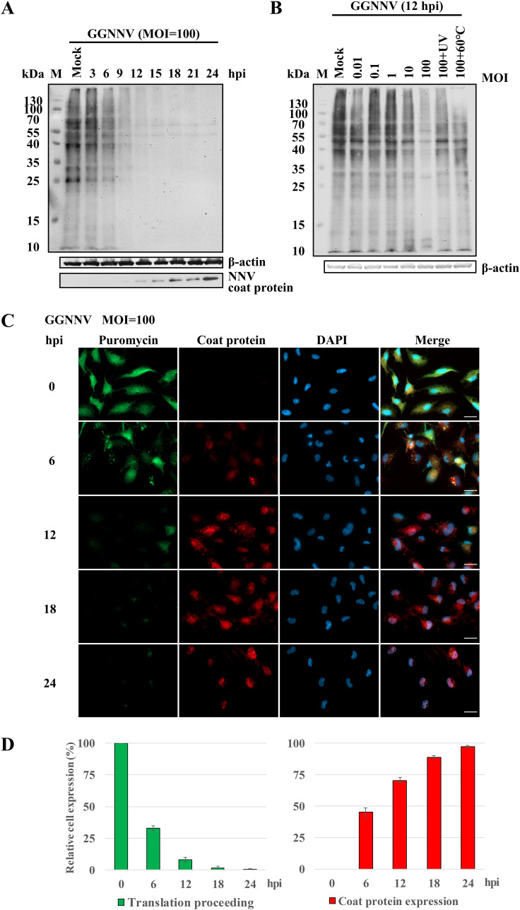
FIG 1.
NNV infection inhibits host translation. Western blot and immunocytochemical staining of puromycin incorporation were performed to evaluate the influence of NNV infection on host translation activity. Puromycin mimics aminoacyl tRNA and is incorporated into newly synthesized proteins during translation. Thus, it can serve as an indicator of translation. Host translation shutoff was observed with the puromycin-label (SUnSET) method after giant grouper nervous necrosis virus (GGNNV) infection in grouper brain (GB) cells. (A and B) GB cells were infected with GGNNV (multiplicity of infection [MOI] = 100) for different time periods (A) or for 12 h with different dosages of GGNNV (B). Puromycin (20 μg/ml) was added to the medium, and the cells were incubated at 28°C for 1 h. As a control, GGNNV was treated with heat (60°C, 1 h) or UV (0.24 J/cm2). The cells were lysed, and lysates were used for SDS-PAGE and immunoblotting analysis. The puromycin incorporated into newly synthesized proteins was detected using an anti-puromycin antibody. A β-actin immunoblot is shown as a loading control. A monoclonal antibody (RG-M18) against NNV coat protein was used to detect the GGNNV level (56). (C) Newly synthesized proteins in GGNNV-infected GB cells were detected by immunocytochemistry. GB cells were infected with GGNNV at an MOI of 100. At 0, 6, 12, 18, and 24 h postinfection (hpi), cells were harvested for immunocytochemistry. Puromycin was added to the medium of infected cells for 1 h prior to harvest. The GGNNV-infected GB cells were washed twice with PBS and then fixed in 4% paraformaldehyde, permeabilized in 0.25% Triton X-100, and processed for immunocytochemistry. Mouse monoclonal antibody RG-M18 was used to detect GGNNV coat protein (red). Newly synthesized proteins with incorporated puromycin were detected with a mouse anti-puromycin Alexa Fluor 488-conjugated monoclonal antibody (green). Nuclei were stained with DAPI (blue). Bar = 20 μm. (D) Statistical analysis of the relative cell expression levels in puromycin-labeling (green) and NNV coat protein expression (red) cells of the immunocytochemical experiment. The means ± standard deviation from three independent experiments are plotted. M, protein molecular weight marker.

