FIG 3.
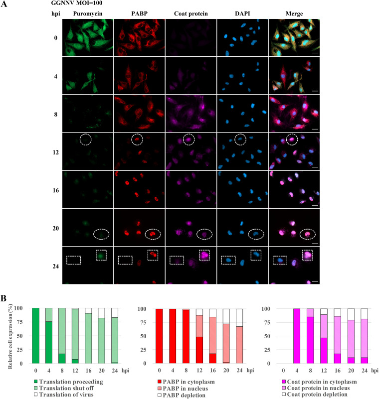
Effects of NNV infection on the expression of newly synthesized proteins, PABP, and viral coat protein. (A) The effect of GGNNV infection on expression of newly synthesized proteins. PABP and coat protein in GB cells were examined by immunostaining. A total of 2 × 105 GB cells were seeded on cover glass in a 3-cm culture dish 15 h before GGNNV infection (MOI = 100). Puromycin (20 μg/ml) was added to the medium of infected or uninfected cells 1 h before the cells were harvested for immunocytochemical staining at the indicated times. Newly synthesized proteins with incorporated puromycin were detected using a mouse anti-puromycin Alexa Fluor 488-conjugated monoclonal antibody (green). PABP was detected with a rabbit anti-PABP polyclonal antibody (red). Mouse monoclonal antibody RG-M18 was used to detect GGNNV coat protein (violet). Nuclei were stained with DAPI (blue). White circles indicate cells with a low level of newly synthesized protein and nuclear localization of both PABP and NNV coat protein. White ovals indicate cells with a low level of newly synthesized protein and nuclear localization of PABP but NNV coat protein depletion. White squares indicate cells with newly synthesized viral protein and nuclear localization of PABP as well as nuclear/cytoplasmic localization of NNV coat protein. White rectangles indicate cells with a low level of newly synthesized protein and both PABP and NNV coat protein depletion. Bar = 20 μm. (B) Statistical analysis of the relative cell expression levels in puromycin-labeling (green), PABP expression (red), and NNV coat protein expression (violet) cells of immunocytochemistry. Cell number, n = 65 to 104 cells analyzed for each time point. GGNNV, giant grouper nervous necrosis virus; hpi, hours postinfection; MOI, multiplicity of infection; PABP, polyadenylate binding protein.

