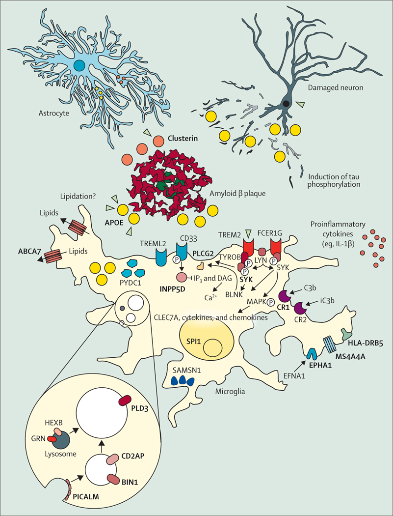
Figure 4: The cellular phase of Alzheimer’s disease.
Although amyloid plaques (red, middle of the figure) and tau phosphorylation and tangles (neurons, top right corner) are still considered the defining features of Alzheimer’s disease, the focus of research has been widened from neurons to the response of other cell populations in the disease.71 The microglia-mediated inflammation, known for decades to be present in Alzheimer’s disease,72 has finally taken centre-stage in functional research on the pathogenesis of the disease. Many of the risk-genes protein products (bold and capitals) identified in Alzheimer’s disease (figure 3) are expressed and have functions in microglia. These genes become upregulated when microglia are exposed to amyloid plaques and many of the Alzheimer’s disease risk genes are enriched in the disease-associated microglia response that characterises this cell state.73–75 Other genes involved in this response and moderately positive in genome-wide association studies are indicated as well. Adapted from Sierksma et al,76 by permission of EMBO Molecular Medicine.

