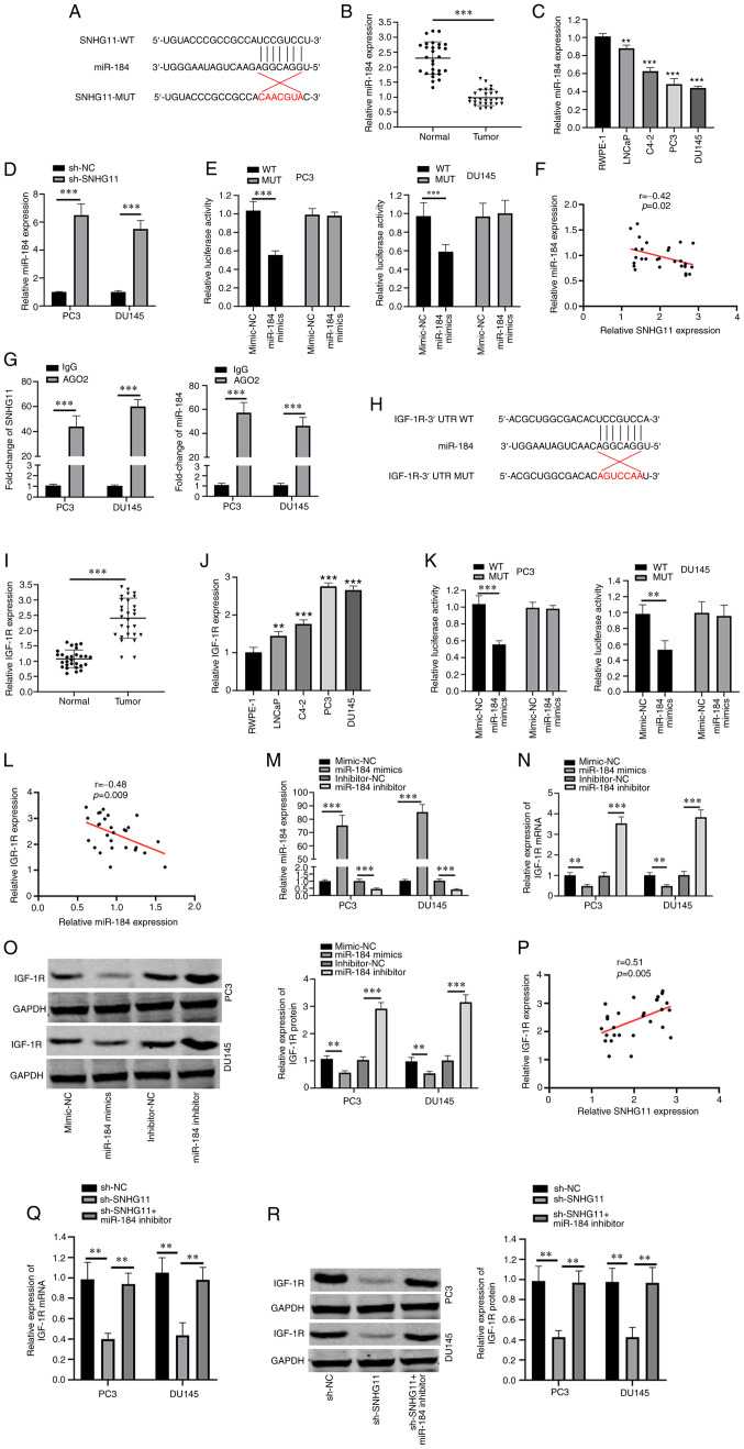
Figure 2.
SNHG11 acts as a sponge for miR-495. (A) Predicted binding site with miR-184 in SNHG11 through bioinformatics analysis. (B) miR-184 expression levels were markedly reduced in PCa tissues compared with that in normal tissues. (C) miR-184 expression levels were markedly decreased in PCa cell lines (LNCaP, C4-2, PC3, DU145) compared with RWPE-1 cell. (D) Silencing SNHG11 increased miR-184 relative expression levels in PC3 and DU145 cells. (E) Luciferase reporter assay revealed that miR-184 mimics inhibited the SNHG20-WT activity in PC3 and DU145 cells. (F) SNHG11 expression negatively correlated with miR-184 expression in PCa tissues. (G) RIP assay results suggested the enrichment of SNHG11 and miR-184 in Ago2-containing beads. (H) Predicted binding sites between miR-184 and IGF-1R through bioinformatics analysis. (I) IGF-1R levels in PCa tissues were significantly increased in comparison with normal tissues. (J) IGF-1R expression was significantly increased in PCa cell lines (LNCaP, C4-2, PC3, DU145), as compared with RWPE-1 cell. (K) miR-184 mimics significantly reduced IGF-1R-WT luciferase activity in PC3 and DU145 cells. (L) IGF-1R expression was negatively associated with miR-184 expression in PCa tissues. (M) Effects of miR-184 mimics or miR-184 inhibitor on miR-184 expression in PC3 and DU145 cells as detected using RT-qPCR. (N and O) miR-184 markedly reduced IGF-1R mRNA and protein levels in PC3 and DU145 cells; however, this was reversed with the use of miR-184 inhibitor. (P) SNHG11 expression positively correlated with IGF-1R expression in PCa tissues. (Q and R) SNHG11 knockdown inhibited the IGF-1R mRNA and protein expression levels, and these effects were reversed with the addition of the miR-184 inhibitor in PC3 and DU145 cells. **P<0.01 and ***P<0.001, in comparison with the control group. SNHG11, lncRNA small nucleolar RNA host gene 11; PCa, prostate cancer; IGF-R1, insulin-like growth factor 1 receptor.

