FIG 1.

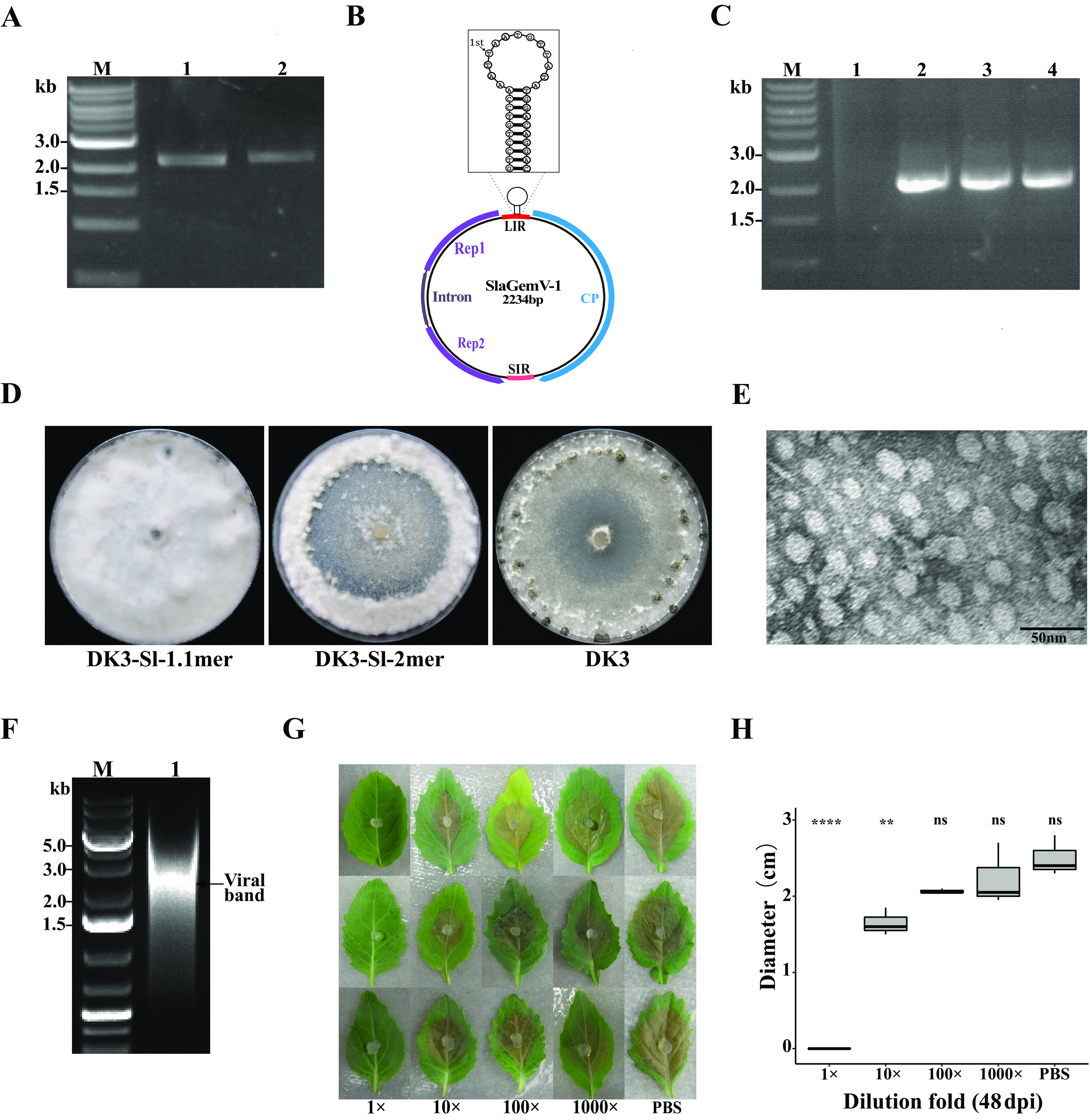
SlaGemV-1 replication in Sclerotinia sclerotiorum after transfection with infectious clones was confirmed. (A) PCR amplification from extracted mycelial DNA of SlaGemV-1-transfected S. sclerotiorum using primers pJetR and 3′R. Lanes: M, 1-kb ladder; 1, 1.1-mer transfection; 2, 2-mer transfection. (B) SlaGemV-1 genome organization with the nonanucleotide structure specified. (C) PCR amplification with primers pJetR and 3′R and with the rolling circle amplification product from the transfected S. sclerotiorum mycelial DNA as the template. The resulting PCR products were subjected to Sanger sequencing to confirm their identities. Lanes: M, 1-kb ladder; 1, RCA product with a smear; 2, PCR product using the RCA product from the 1.1-mer-transfected culture as the template; 3, PCR product of RCA product from 2-mer-transfected culture; 4, positive control from PCR product without RCA from the transfected culture. (D) Comparison of virus-transfected S. sclerotiorum (strain DK3) grown on PDA for 21 days after transfection with pJet-SlaGemV-1 1.1-mer and 2-mer constructs. (Left) pJet-SlaGemV-1 1.1-mer transfection; (middle) pJet-SlaGemV-1-2-mer transfection; (right) original DK3 strain, used for comparison. (E) Transmission electron microscopy image of purified SlaGemV-1 particles. (F) Agarose gel (1%) electrophoresis of DNA extracted from 25% sucrose gradient fractions containing virus particles. (G) Viral particles (1 mg/ml) extracted from infected S. sclerotiorum mycelia were diluted to different concentrations (1 mg/ml, 0.1 mg/ml, 0.01 mg/ml, and 0.001 mg/ml; PBS, phosphate-buffered saline control) used as inoculants (10 µl) on virus free agar plug to inhibit fungal growth. As the viral particles were resuspended in PBS, all dilution used PBS. Each row represents one replication for three replications shown here. (H) Dose-response relationship of undiluted and diluted treatments of viral particles compared to the PBS control. Two-way analysis of variance and Bonferroni post hoc test. **, P < 0.01; ****, P <0.0001.
