
Figure 10.
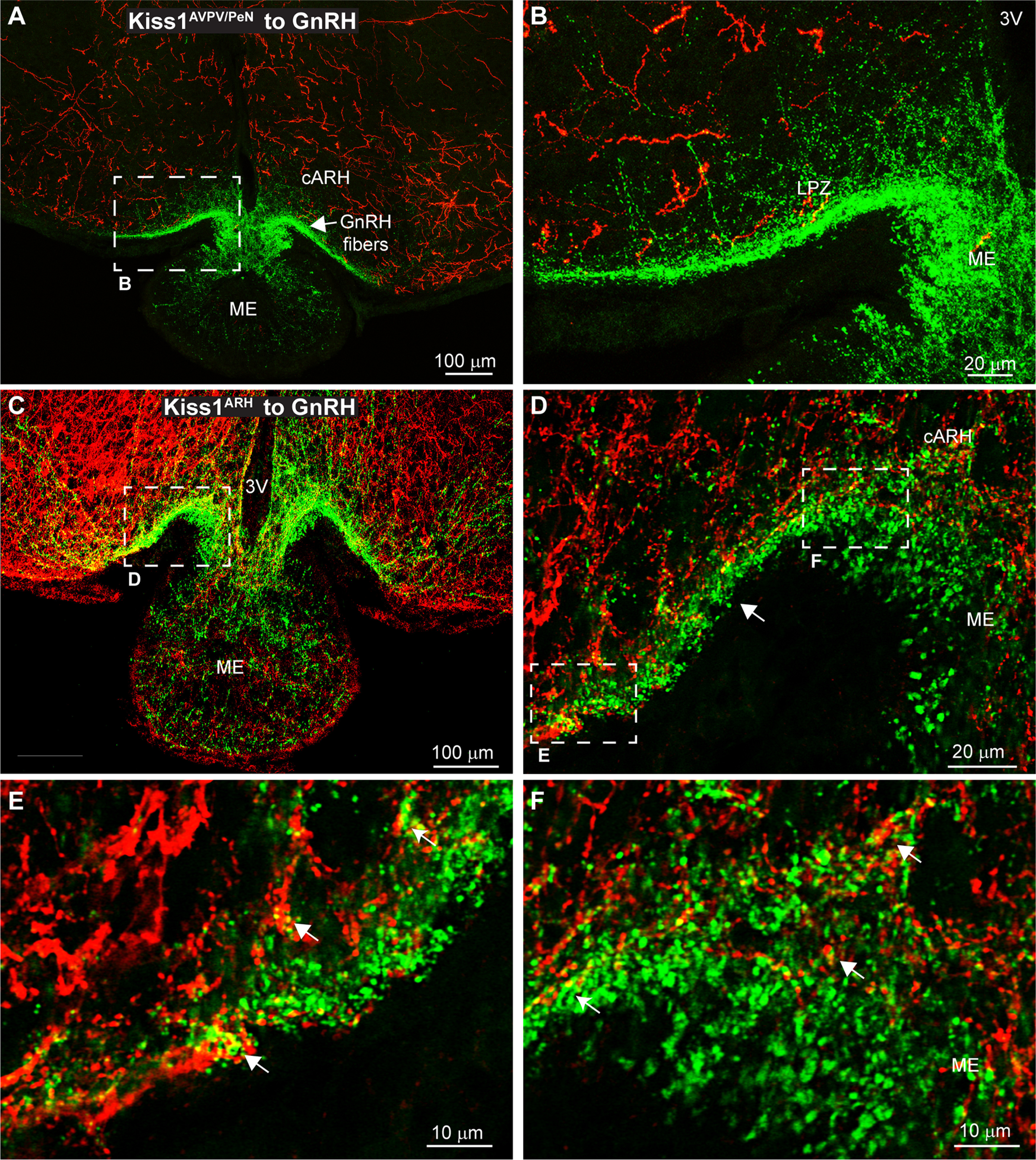
Interactions between GnRH and Kiss1 fibers in the ME. Confocal image montages of double label ICC that show Kiss1 fibers (ChR2-mCherry) originating from either the AVPV/PeN or ARH. GnRH fibers (green) enter the ARH laterally and along the 3V from the POA before entering the ME. A, Kiss1AVPV/Pen fibers are diffuse in the ARH and do not enter the ME. B, Expanded view of the dashed box from A. Few Kiss1AVPV/PeN fibers reach the ventral surface where GnRH fibers concentrate, which would suggest interactions are unlikely. C, Confocal image montage displays staining of Kiss1ARH cell bodies and projections in the cARH and ME. D, Single optical slice (1 μm thick) of the region outlined in C. GnRH fibers enter the ARH laterally (left) to briefly form a bundle (white arrow) that runs along the ventral surface before dispersing in the ME. D, Expanded view of the left white box in C. The occasional presence of yellow pixels and lack of black pixels between Kiss1ARH and GnRH fibers suggests the presence of interactions. E, F, Expanded view of the regions from the white boxes in D. As the GnRH fibers enter the ME the lack of black pixels between Kiss1ARH::ChR2-mCherry and GnRH fibers and presence of yellow pixels suggest close contacts (white arrows) between fibers.