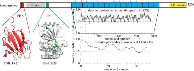
Figure 1.
Schematic diagram of human Ki-67. This schematic of Ki-67 isoform I highlights conserved regions. The Ki-67 forkhead associated (FHA) domain (red) is accompanied by its solution NMR structure (PDB:1R21) [37]. Exon 7, highlighted in grey, is missing from isoform II. The PP1-binding domain (green) is accompanied by the crystal structure of the Ki-67 (green):PP1γ (grey) holoenzyme complex (PDB:5J28) [35]. The repeat region is highlighted in blue with each individual repeat marked. The FKELF motif, which binds the original Ki-67 monoclonal antibody, is indicated by an asterisk. The disorder probability graphs show disorder across all (top) and the first (bottom) repeat calculated by the PrDOS software [38]. The red line indicates a disorder probability of 0.5. Anything above this is highly likely to be disordered. The LR domain (yellow) is responsible for DNA binding and chromosome association of Ki-67.

