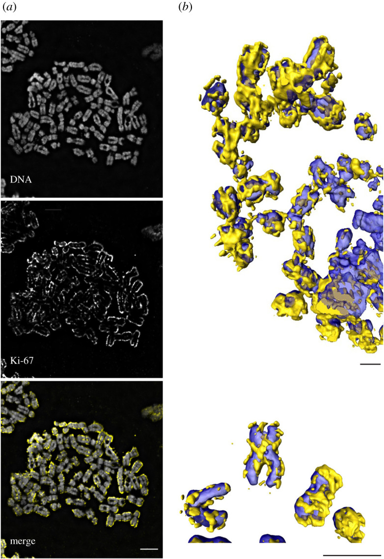
Figure 6.

Ki67 is enriched at the chromosome periphery during mitosis. (a) Immunofluorescence microscopy of chromosome spread from HeLa CDK1as cells, showing Ki-67 (yellow) enriched on the surface of the DNA (grey). (b) Selected chromosomes from a chromosome spread similar to that in A were rendered in three-dimensions using AMIRA, showing how Ki-67 coats the chromosome surface. The discontinuous appearance of the layer is likely to be an artefact of the rendering process in AMIRA. HeLa CDK1as [85] were maintained in DMEM (Invitrogen) supplemented with 5% fetal bovine serum (Invitrogen) and 100 U/ml penicillin G and 100 µg ml−1 streptomycin sulphate (Invitrogen). Colcemid was added to the culture at a final concentration of 0.1 µg ml−1 for 75 min before harvesting by mitotic shake off. Cells were treated with hypotonic solution (75 mM KCl) for 10 min before Cytospin at 1900 RPM, High intensity for 10 min. Following fixation with 4% paraformaldehyde for 10 min at 37°C, immunofluorescence of metaphase chromosomes was carried out using Anti-Ki-67 mouse monoclonal antibody 1 : 500 (9449S, Cell Signalling) and Alexa 555 1 : 1000. Chromosomes were mounted with VECTASHIELD Antifade Mounting Medium with DAPI. Scale bar, 5 mm.
