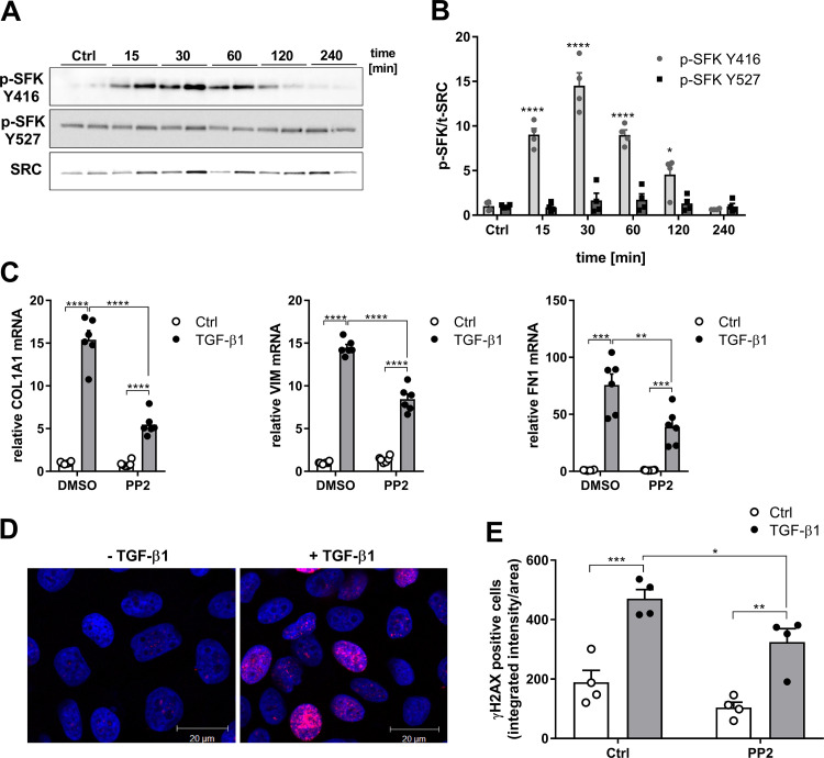
Figure 2.
TGF-β1 induces SFK-mediated profibrotic gene expression and DNA damage response. A: representative Western blots indicating phosphorylation of SRC kinases at tyrosine 416 and 527 after stimulation with TGF-β1 (10 ng/mL). B: densitometry analysis of 4 replicates from 2 independent experiments. C: gene expression of COL1A1, VIM, and FN1 after stimulation with TGF-β1 (10 ng/mL) for 24 h, in the absence or presence of PP2, determined by RT-PCR (n = 6). D: representative immunofluorescence imaging of γH2AX (red) and 4′-6-diamidino-2-phenylindole dihydrochloride (DAPI; blue) in unstimulated or TGF-β1–stimulated (4 h) HBE1 cells. E: quantitative analysis of nuclear γH2AX intensity in HBE1 cells in the absence or presence of PP2 (1 µM) with and without TGF-β1 (10 ng/mL) stimulation for 4 h. For each experimental condition, a minimum of 200 cells from a total of 2 independent experiments with 2 replicates were analyzed. Results are expressed as means ± SE (*P < 0.05, **P < 0.01, ***P < 0.001, ****P < 0.0001).

