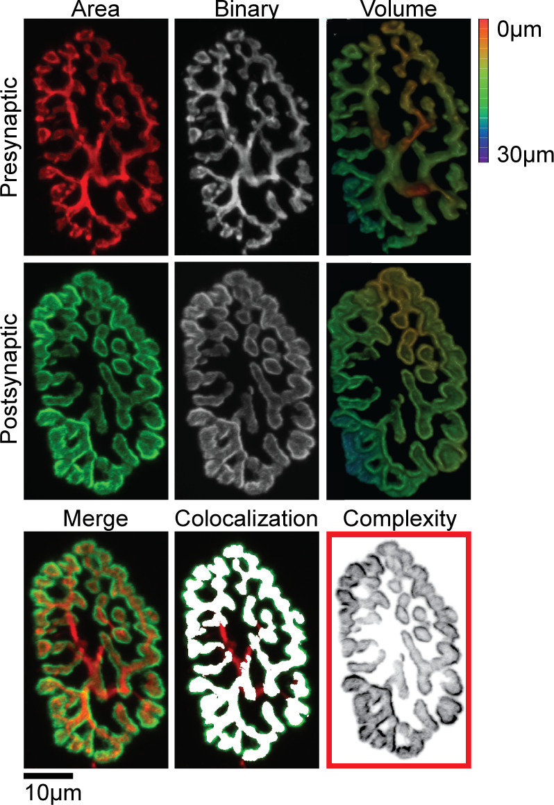
Figure 1.

Individual presynaptic (red) and postsynaptic (green) NMJ structures were stained, imaged, binarized, and used to quantitatively measure two-dimensional (2-D) planar area, three-dimensional (3-D) volume, colocalization, and complexity. Images were also used for visual analysis of NMJ denervation and the appearance of irregular morphological characteristics. Scale bar 10 µm and 3-D color depth scale range 0–30 µm. NMJ, neuromuscular junctions.
