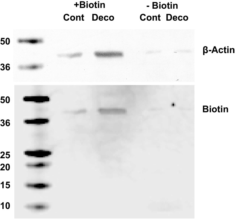
Figure 4.
Biotinylated vs. nonbiotinylated MPs separation: MPs from control and decompressed male mice were isolated, biotinylated, and then lysed. Samples were incubated with magnetic streptavidin beads as described in methods and passed through a magnet to separate biotinylated (shown as +Biotin) from nonbiotinylated proteins (shown as −Biotin). Protein from 165,000 MPs was loaded into each lane for SDS-PAGE. Western blots probed for β-actin and biotin are shown. Probing for IL-1β did not demonstrate bands (not shown). Molecular weight standards (in kDa) are shown at left. IL-1β, interleukin-1β; MP, microparticles.

