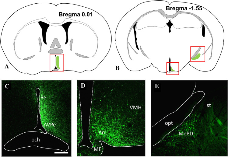
Fig. 4.
Illustration of the brain areas containing Cre expressing KP neurons (A and B), which underwent a virus-based identification in the preoptic (C), arcuate (D), and medial amygdala (E) regions, respectively. The expressed YFP has been immunohistochemically amplified to show membranes of KP neurons. Modifications of the atlas images from the mouse brain atlas of Paxinos and Franklin’s (2012). Scale bar 200 µm. Arc arcuate nucleus, AVPe anteroventral periventricular nucleus, ME median eminence, MePD posterodorsal subdivision of medial amygdala, och optic chiasm, opt optic tract, Pe periventricular hypothalamic nucleus, st stria terminalis, VMH ventromedial hypothalamic nucleus

