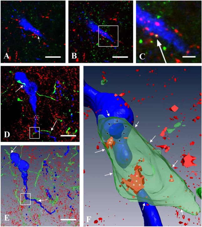
Fig. 5.

CB1-immunoreactivity in KP afferents of GnRH neurons in the medial preoptic area of OVX-EB mice. Demonstration of appearance of CB1-immunoreactivity in one of the KP-IR afferent fiber (white arrow) in confocal microscopic Z-stack series. A A single optical slice shows multiple KP-immunoreactive (IR) varicosities (red and white arrows) in apposition to a GnRH-IR neuron (blue). B is an adjacent optical slice. The boxed area in this image is magnified in C to demonstrate the yellow-colored double-labeled KP-IR varicosity (white arrow). D Yellow fluorescent protein (YFP)-positive, green-colored axon varicosities (KP fibers after viral and immunohistochemical detection) in apposition to two adjacent gonadotropin-releasing hormone (GnRH)-IR cells (blue) shown in merged three optical slices. E The 3D rendered view of all optical slices of the same structures. The GnRH neurons are embedded in a tissue showing punctate CB1-IR sites (red), where CB1-IR clusters (dotted circles) and KP-IR fibers (white arrows) are in association with GnRH-IR cell bodies or processes. F The projection image of the 3D reconstructed area of E (white rectangle) is shown at higher power. Some of the CB1-IR sites (white asterisks) are visible only, if the KP-IR fiber is made semi-transparent. The CB1-IR sites in the membrane of the KP-IR fiber (white arrows) turn up in orange-red color marking co-localization. The contact site between the KP fiber and the GnRH dendron is marked by black arrowheads. Scale bar 10 µm (on A, B, D, E) and 5 µm (on C)
