Fig. 3.

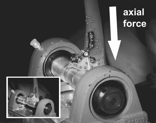
Example of a simulated coronoid fracture after osteosynthetic treatment using an anatomical plate. The fracture fragment was placed on a cylindrical tube. Displacements were measured using a point-based 3D-motion analysis detection system

Example of a simulated coronoid fracture after osteosynthetic treatment using an anatomical plate. The fracture fragment was placed on a cylindrical tube. Displacements were measured using a point-based 3D-motion analysis detection system