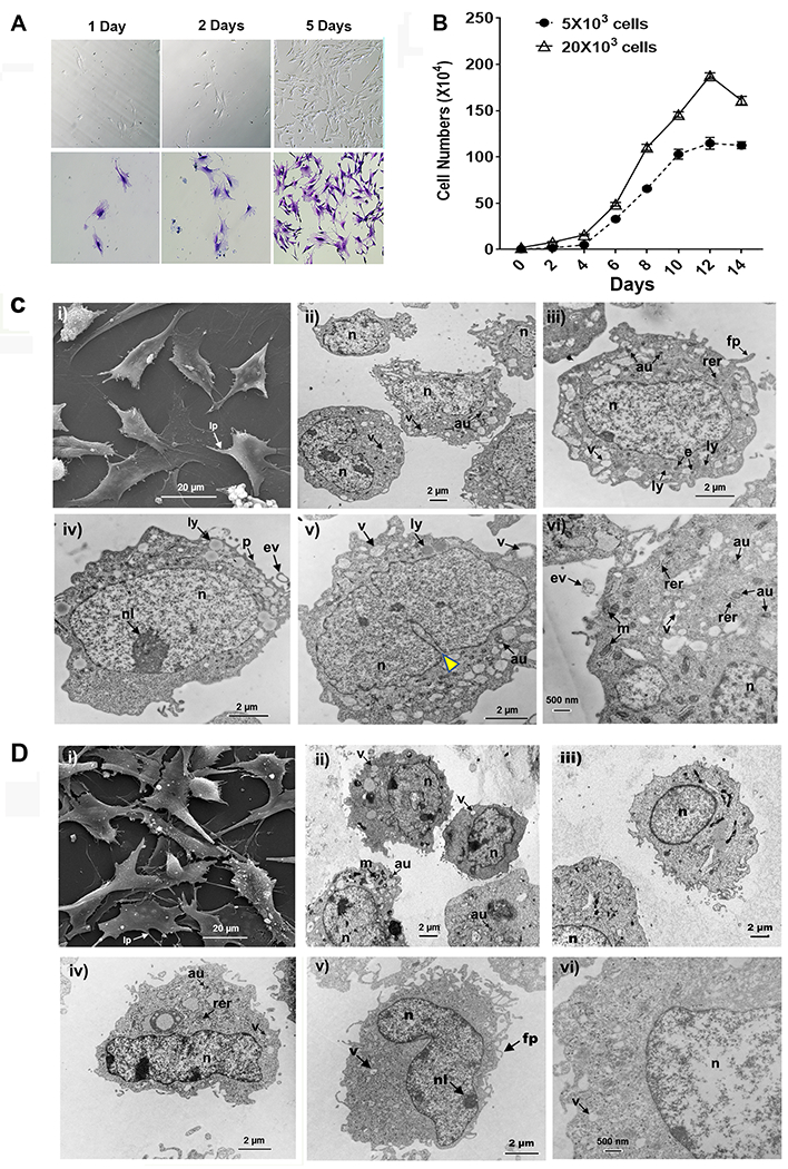
FIGURE 1. Morphology and ultrastructural features of Sca1+ LMSCs.

(A) Sca-1+ LMSCs (passage 6) grown on plastic at low density in CMEM for 1 day, 2 days, and 5 days. Adherent cells were stained with methylene blue. Images of the plastic adherent cells (A, upper panel) and methylene blue-stained cells (A, bottom panel) were taken using a conventional optical microscope. (B) Growth curves of Sca-1+ LMSCs. Results are the mean ± SEM from 2 independent experiments. (C) Scanning electron micrograph (i) of Sca-1+ LMSCs grown on a plastic surface (Theranox™ plastic coverslips) for 5 days. Note the flattened cell with lamellipodia and many thin filopodia as well as the fibroblast-like morphology. Magnification, x3000. Scale bar = 20 μm. Transmission electron micrographs (ii – vi) of cultured Sca-1+ LMSCs at (ii) magnification x8000, scale bar = 2 μm, (iii, iv, v) magnification x12,000, scale bars = 2 μm, and (vi) magnification x20,000, scale bar = 500 nm. D) Scanning electron micrograph (i) of 3T3 MEFs grown on a plastic surface (Theranox™ plastic coverslips) for 5 days. Note the flattened cells with processes spreading outward. Magnification, x2500, scale bar = 20 μm. Transmission electron micrographs (ii – vi) of 3T3 MEFs grown in CMEM for 5 days (ii) magnification x8000, (iii) magnification x12,000, (iv) magnification x12,000, (v) magnification x12,000, scale bars = 2 μm, and (vi) magnification x20,000, scale bar = 500 nm. au, autophagosome; e, endosome; ev, extracellular vesicle; fp, filopodia; lp, lamellipodia; ly, lysosome; m, mitochondria; n, nucleus; nl, nucleoli; p, plasma membrane; rer, rough endoplasmic reticulum; v, vesicle; yellow Δ, invagination of nuclear envelope.
