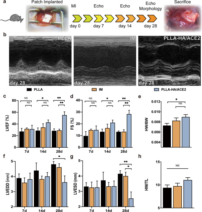
Fig. 6. rhACE2 patch preserve left ventricular function after acute myocardial infarction in vivo.
a The animal research timeline design with images represented the acute MI model and successful implantation of rhACE2 patch. Echo, echocardiography. b Representative M-mode parasternal long axis view of left ventricle echocardiographic images of different groups at day 28 after LAD coronary artery ligation. Statistical analysis of left ventricular ejection fraction (c), shortening fraction (d), heart weight/body weight ratio (e), LV end-diastolic diameter (f), LV end-systolic diameter (g) and heart weight/tibial length (h) determined by echocardiography obtained from PLLA, intramyocardial injection (IM) and PLLA-HA/ACE2 treatment group at day 7, day 14 and day 28 after operation. n = 5/group. Data were represented as the mean ± SEM and analyzed for statistical significance using two-way ANOVA followed by Tukey’s multiple comparison test; NS no significant difference, *P < 0.05, **P < 0.01.

