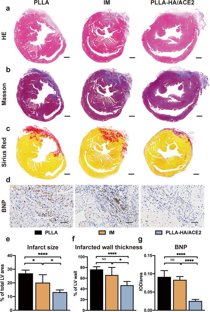
Fig. 7. Histological analysis of rhACE2 patch effect on MI-induced cardiac remodeling.
H&E stained section (a), Masson’s trichrome staining (b) and Sirius Red staining (c) of mice hearts at low magnification after heart was obtained 28 days after MI. Scale bars represented 500 μm. d Brain natriuretic peptide (BNP) immunohistochemical staining of hearts from PLLA, intramyocardial injection (IM) and PLLA-HA/ACE2 treatment group 28 days after MI. Scale bars represented 40 μm. Morphometric parameters including the percentage of infarct size percentage of total LV area (e) and the percentage of infarcted thickness of LV anterior wall (f) were measured from the Sirius Red stained slides via ImageJ software. n = 5/group. g The integrated optical density (IOD)/area ratios of BNP were quantified from the immunohistochemical stain by Image-Pro Plus software. n = 5/group. Data were represented as the mean ± SEM and was analyzed for statistical significance using one-way ANOVA followed by Tukey’s multiple comparison test; NS no significant difference, *P < 0.05, ****P < 0.0001.

