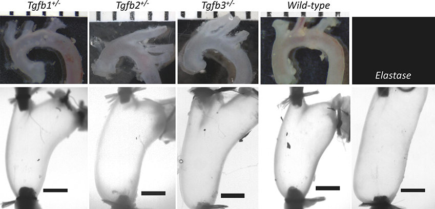
Figure 2.
Representative images of the ascending thoracic aortas from Tgfb1+/−, Tgfb2+/−, Tgfb3+/−, wild-type, and Elastase infused wild-type mice prior to, (top) and following cannulation, axial extension, and pressurization to 100 mmHg (bottom). (top) Ruler with mm units and (bottom) 1 mm scale bars. Elastase infusion is performed in the loaded configuration.

