
Figure 3: Full length Meikin and the C-Meikin cleavage fragment bind Plk1 and promote Rec8 cleavage similarly.
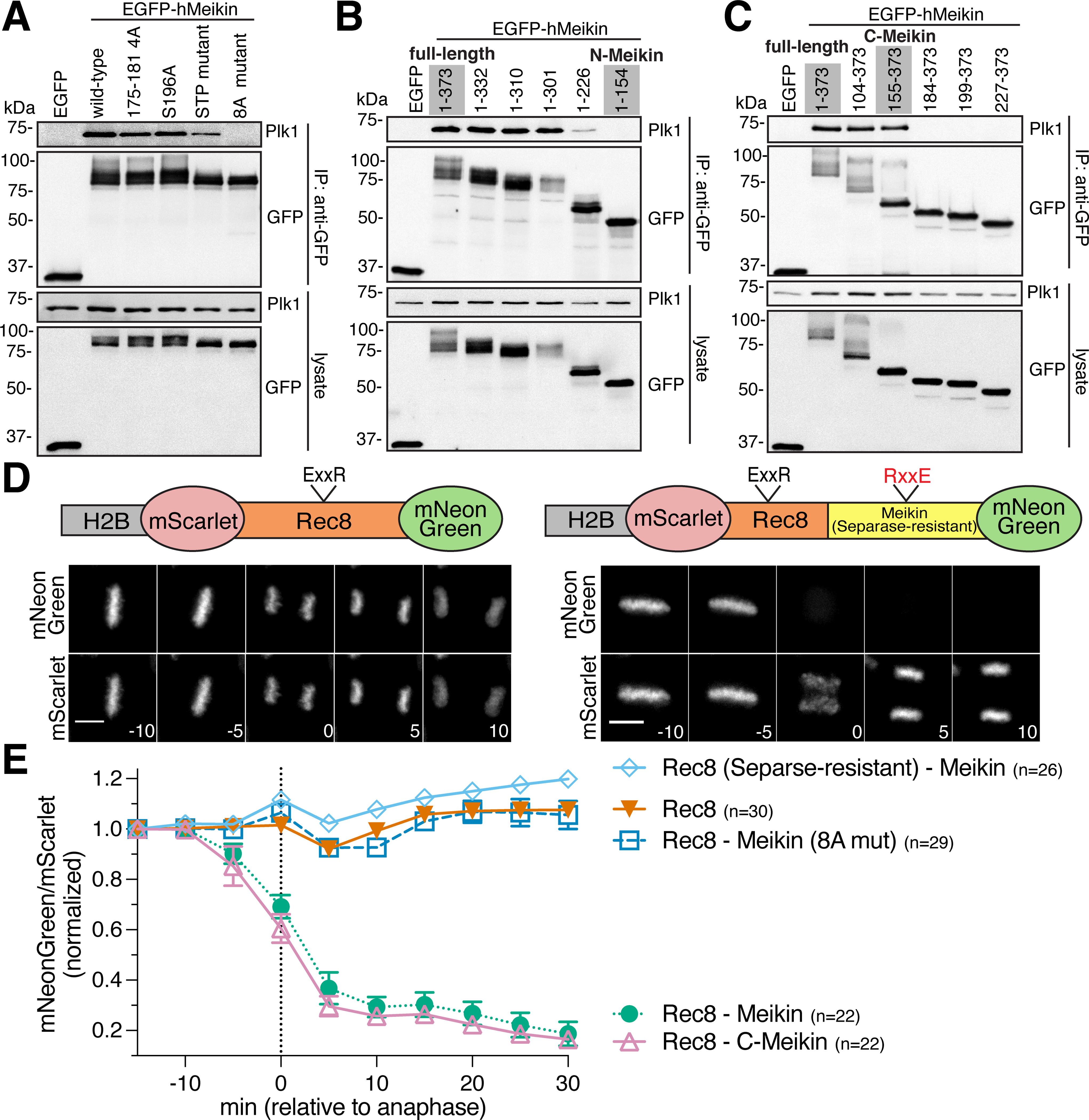
A. Induced HeLa cells were arrested with STLC. GFP-immunoprecipitates were analyzed by Western blot. Specific residues changed to alanine in each mutant are described in the Key Resources Table. B and C. Western blot analysis of GFP immunoprecipitates from cells expressing hMeikin constructs as above. D. Time-lapse montage of HeLa cells expressing the indicated Rec8 cleavage sensor. The sensor contains a fragment of hRec8 including the predicted Separase cleavage sites. Numbers indicate minutes relative to anaphase onset. Scale bars, 10 μm. E. Quantification of Rec8 cleavage sensor analyzed and represented as in Figure 1. n = number of mitoses analyzed. See also Figure S3 and S4.