
Figure 4: Separase cleavage of Meikin is required for proper meiosis II chromosome alignment.
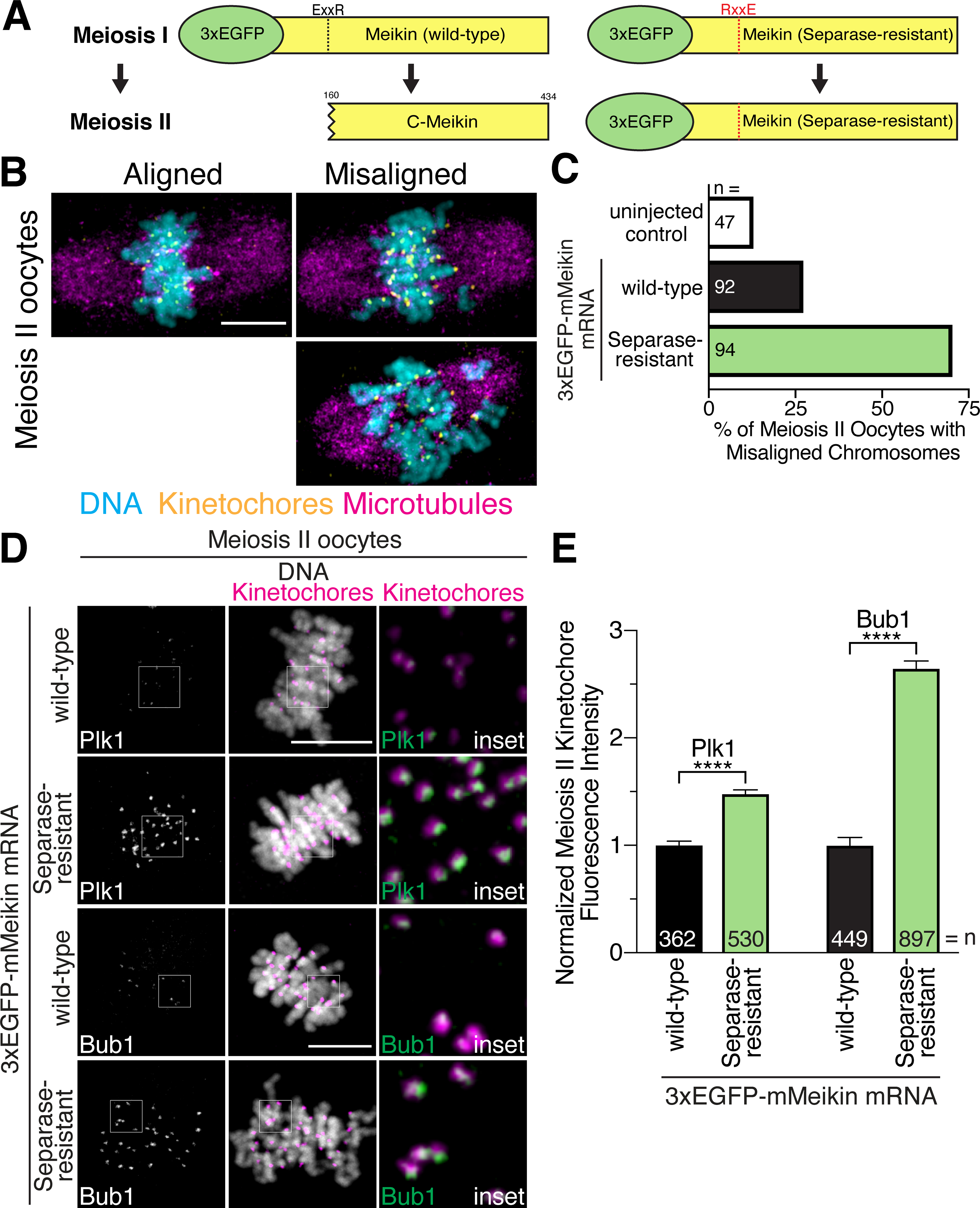
A. Schematic of mMeikin constructs and their expected behaviors at meiosis II. B. Representative immunofluorescence images of chromosome misalignment defects observed in meiosis II oocytes. C. Quantification of defects observed in injected oocytes. n = number of oocytes analyzed. D. Mouse oocytes injected with the indicated Meikin mRNA and stained for Plk1 or Bub1 at meiosis II. Images of similarly stained cells are scaled identically. E. Quantification of kinetochore intensity of Plk1 and Bub1 in meiosis II oocytes injected with the indicated mMeikin mRNA. Values from two experiments were pooled. n = number of kinetochores analyzed. Means and 95% confidence intervals are presented. ****P < 0.0001, two-tailed t-test. Kinetochores are stained with mouse CENP-C antibody. Scale bars, 10 μm. Insets, 5 μm. See also Figure S5.