
Figure 5: The C-Meikin cleavage fragment is required for meiosis II chromosome alignment.
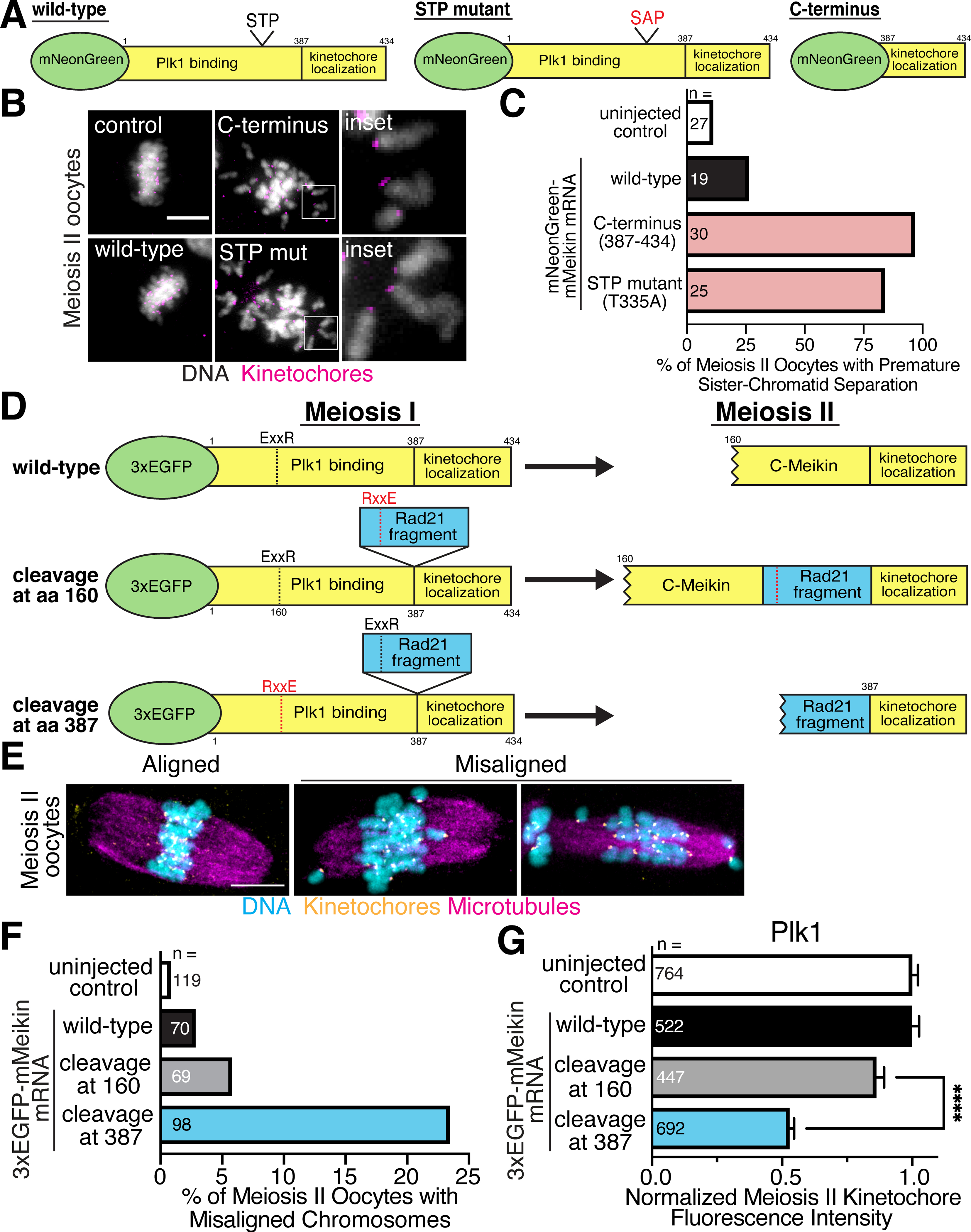
A. Schematic of mMeikin constructs. B. Representative immunofluorescence images of oocytes injected with the indicated Meikin mRNA and matured to meiosis II. Insets show separated chromatids. Kinetochores are stained with Hec1 antibody. C. Quantification of premature sister chromatid separation defects observed in meiosis II oocytes expressing the indicated Meikin mRNA. Kinetochores are stained with Hec1 antibody. D. Schematic of mMeikin constructs and their expected behaviors at meiosis II. A fragment of hRad21 (amino acids 142–275) containing the conserved Separase cleavage site was used. A fragment of hRad21 (amino acids 142–275) was inserted between the Plk1 binding and kinetochore localization domains of Meikin. Charge-swap mutations in the Separase-cleavage sites of Meikin (E156R, R159E) or Rad21 (E169R, R172E) were used to direct the location of Separase cleavage during anaphase I. E. Representative immunofluorescence images of chromosome misalignment defects observed in meiosis II oocytes. Kinetochores are stained with mouse CENP-C antibody. F. Quantification of meiosis II chromosome misalignment defects observed in oocytes injected with the indicated mMeikin construct. n = number of oocytes analyzed. G. Quantification of Plk1 kinetochore intensity in meiosis II oocytes injected with the indicated mMeikin mRNA. Means and 95% confidence intervals from pooled results of two experiments are presented. n = number of kinetochores analyzed. ****P < 0.0001, two-tailed t-test. Scale bars, 10 μm. Insets, 8 μm. See also Figure S6.