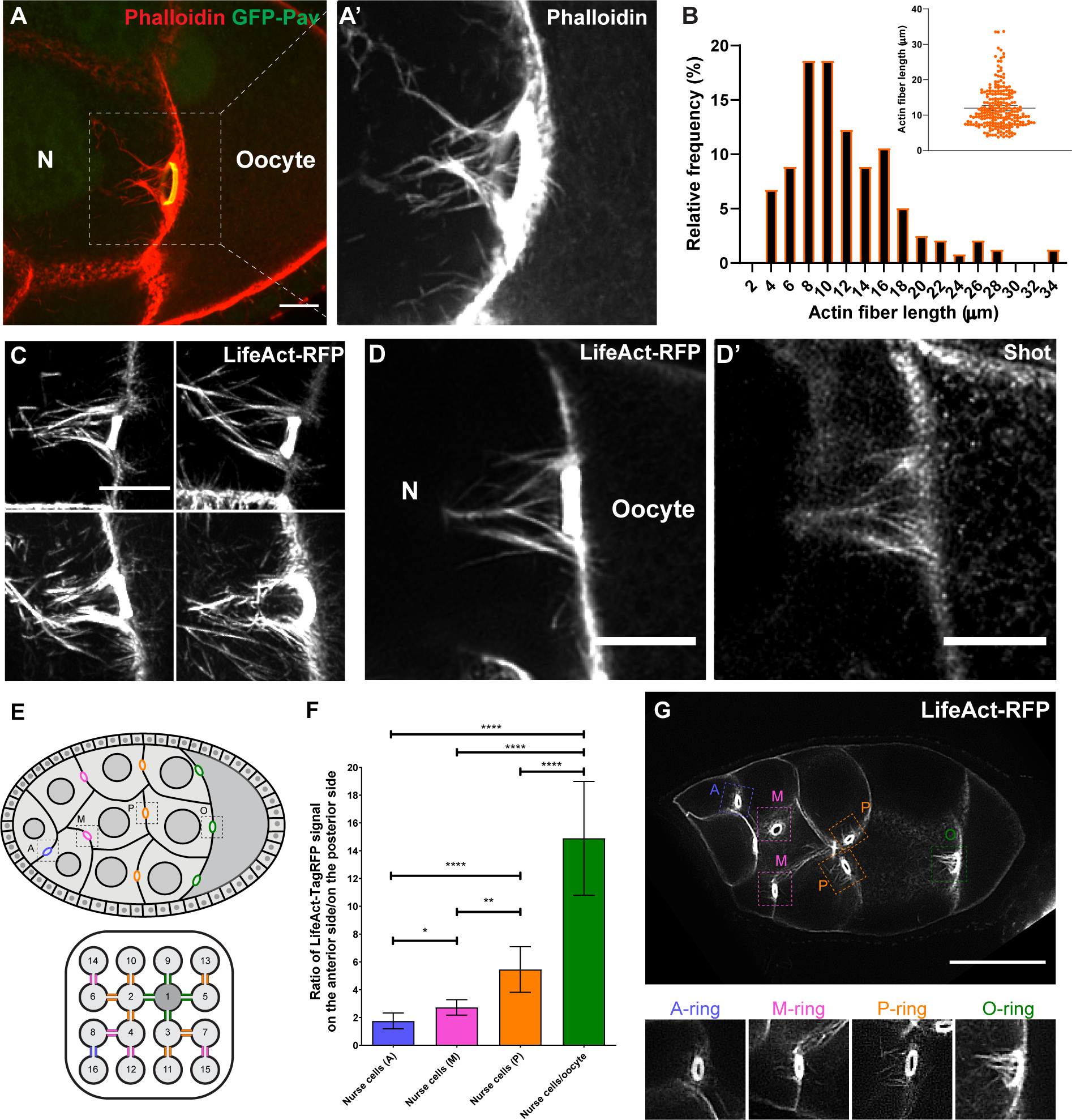
Figure 4. Shot is localized at the asymmetric actin fibers of the nurse cell-oocyte ring canals.

(A-A’) Rhodamine-conjugated phalloidin staining shows asymmetric actin fibers (the white dashed box) at the ring canal (ring canal inner rim is labeled with GFP-Pavarotti) on the nurse cell side, not on the oocyte side.
(B) Quantification of the length of actin fibers on the nurse cell side. The lengths of the four longest actin fibers were measured for each ring canal (59 ring canals from 15 control egg chambers). The average actin fiber length on the nurse cell side is 12.0 ± 0.7 μm (mean ± 95% confidence interval).
(C) Asymmetric actin fibers, labeled with TagRFP-tagged LifeAct, are seen at all four ring canals connecting nurse cells and the oocyte in a live sample. See also Video S8.
(D-D’) A representative image of Shot antibody staining in a TagRFP-LifeAct-expressing egg chamber. Shot is localized at the asymmetric actin fibers on the nurse cell side of the ring canal, but it is not concentrated in the F-actin core of the ring canal inner rim.
(E) Schematic illustrations of a stage 8 Drosophila egg chamber and an interconnected 16-cell germline cyst, including 1 oocyte (cell 1) and 15 nurse cells (cells 2–16, numbered according to the order of cell divisions). Ring canals are categorized depends on their relative distance to the oocyte [62] and are color-coded: (1) nurse cell-oocyte ring canals, directly connected to the oocyte, green, “O”; (2) posterior nurse cell-nurse cell ring canal, having one nurse cell between this ring canal and the oocyte, orange, “P”; (3) middle nurse cell-nurse cell ring canal, having two nurse cells between this ring canal and the oocyte, magenta, “M”; (4) anterior nurse cell-nurse cell ring canal, having three nurse cells between this ring canal and the oocyte, blue, “A”.
(F) The asymmetry of actin fibers is quantified as the ratio of LifeAct-TagRFP fluorescence signal on the anterior side to the signal on the posterior side of the ring canals (see more details in Materials and Methods). Numbers of ring canals for each type: anterior nurse cell-nurse cell ring canals (A), N=17; middle nurse cell-nurse cell ring canals (M), N=46; posterior nurse cell-nurse cell ring canals, N=55; nurse cell- oocyte ring canals (O), N=70. Unpaired t tests with Welch’s correction were performed in following groups: “O” and “P”, p<0.0001 (****); “O” and “M”, p<0.0001 (****); “O” and “A”, p<0.0001 (****); “P” and “M”, p=0.0024 (**); “P” and “A”, p<0.0001 (****); “M” and “A”, p=0.0143 (*).
(G) A representative image of a stage 8 egg chamber expressing LifeAct-TagRFP. Four types of ring canals are highlighted in colored boxes with zoom-in images below.
N, nurse cell; scale bars, 10 μm (A, C, D-D’) and 50 μm (G).
