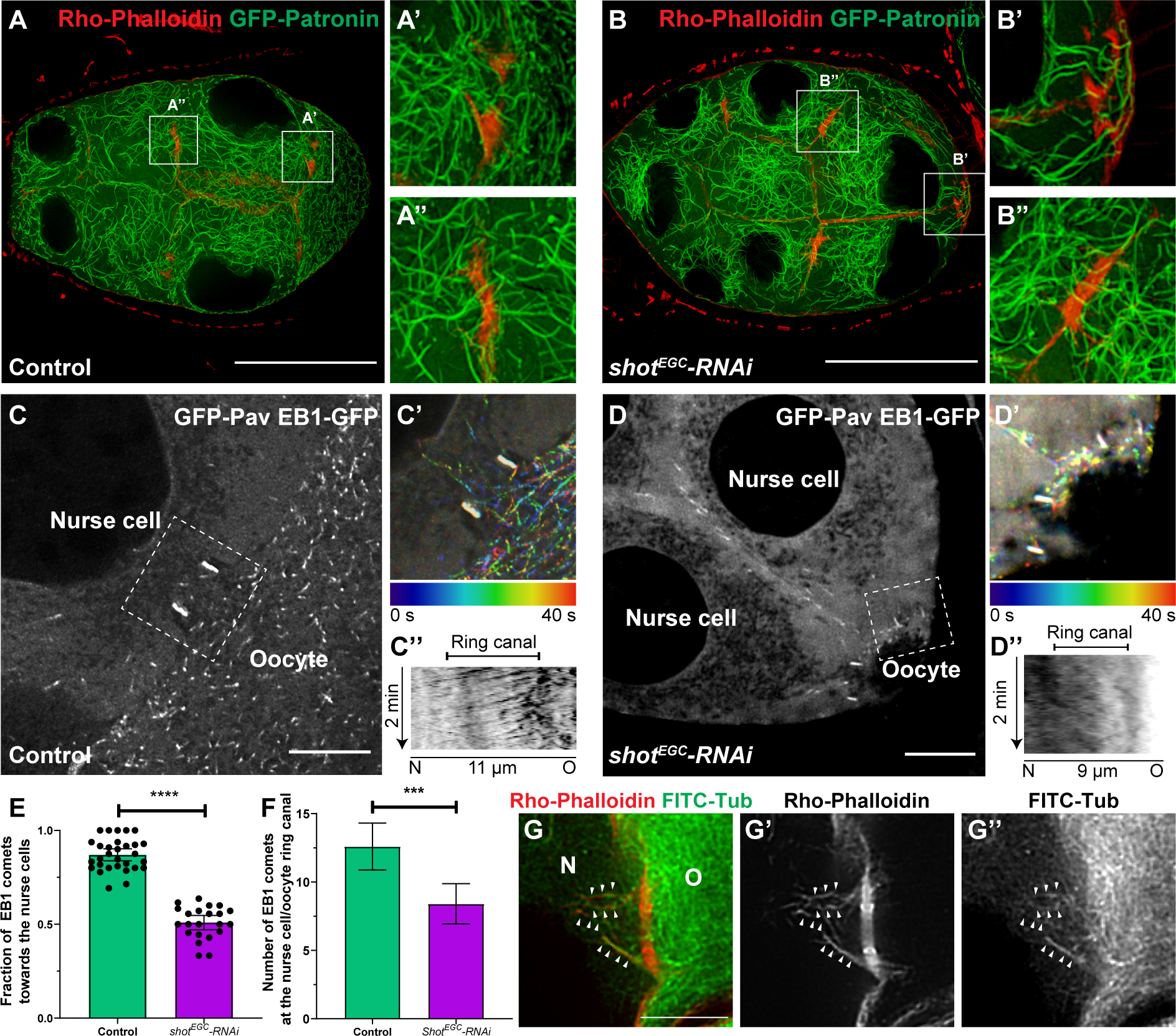
Figure 5. Shot controls microtubule polarity in the nurse cell-oocyte ring canals.

(A-B) Stable microtubule organization is not affected by shot knockdown. (A) In control, stable microtubules are localized at the ring canals between the nurse cell and the oocyte (A’) and between two nurse cells (A”). (B) Knockdown of shot does not change stable microtubule distribution at the ring canals between the nurse cells and the oocyte (B’) and between two nurse cells (B”). Stable microtubules are labeled by overexpressed GFP-tagged Patronin, and ring canals are labeled with Rho-Phalloidin staining. Scale bars, 50 μm.
(C-E) Knockdown of shot results in a mixed orientation of microtubules in the ring canals. (C) EBI-GFP-labeled microtubule +end comets in the ring canal (labeled by GFP-Pav) connecting a nurse cell and an oocyte in control. (C’) A color-coded hyperstack of the EB1 comet movement of (C). (C”) A kymograph of EB1 comet movement at the ring canal (the white dashed box in C) in control. (D) EBI-GFP-labeled microtubule +end comets at the ring canal (labeled by GFP-Pav) connecting two nurse cells and an oocyte in shot-RNAi. (D’) A color-coded hyperstack of the EB1 comet movement of (D). (D”) A kymograph of EB1 comet movement at the ring canal (the white dashed box in D) in shot-RNAi. (E) Quantification of the fraction of EB1 comets moving through the ring canals from the oocyte towards the nurse cells in control and in shot-RNAi. Control, N=30; shotEGC-RNAi, N=22; unpaired t test with Welch’s correction between control and shot-RNAi, p-value < 0.0001(****). (F) Quantification of EB1 comet numbers in the nurse cell-oocyte ring canals in control and shot-RNAi. Control, N=30; shotEGC-RNAi, N=22; unpaired t test with Welch’s correction between control and shot-RNAi, p-value= 0.0004 (***).
(G-G”) A representative image of dual labeling of microtubules (by FITC-conjugated tubulin antibody) and F-actin (by Rho-Phalloidin) at a nurse cell-oocyte ring canal. Alignment of microtubules on asymmetric actin filaments are highlighted by small white arrowheads.
(C-D and G-G’) N, nurse cell; O, oocyte; scale bars, 10μm.
