
Figure 1. Identification of a post-transcriptionally repressive region in the PD-L1 3′UTR.
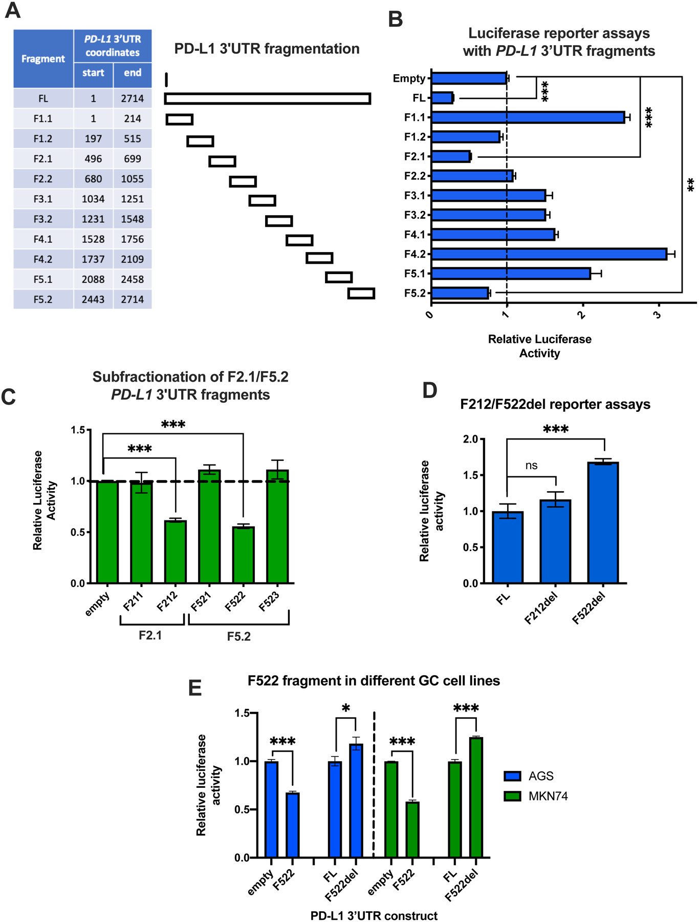
(A) Table with the PD-L1 3′UTR coordinates of the fragments used in the luciferase reporter assays. (B) Luciferase reporter assay with all ten 200–300bp long fragments, along with a full-length (FL) and an empty vector control, in SNU719 cells. The Renilla to Firefly luciferase activity ratio was calculated for each fragment. The results for each fragment were normalized against the empty vector control. (C) Luciferase reporter assays with smaller fractions (100bp-long) of the F2.1 and F5.2 fragments, which demonstrated repressive activity in (B). (D) The F212 and F522 fragments were deleted from the FL PD-L1 3′UTR reporter (del constructs). The activity of the deletion constructs was normalized to the FL control. (E) The same experiment as (C) and (D) for F212 and F522 was repeated in two additional gastric cancer cell lines. The activity of the fragments was normalized to the empty vector control, while the activity of the del reporters was normalized to the FL control. All experiments were performed in triplicate and statistical significance of comparisons was assessed by Student’s t-test. *P < 0.05; **P < 0.01; ***P < 0.001.