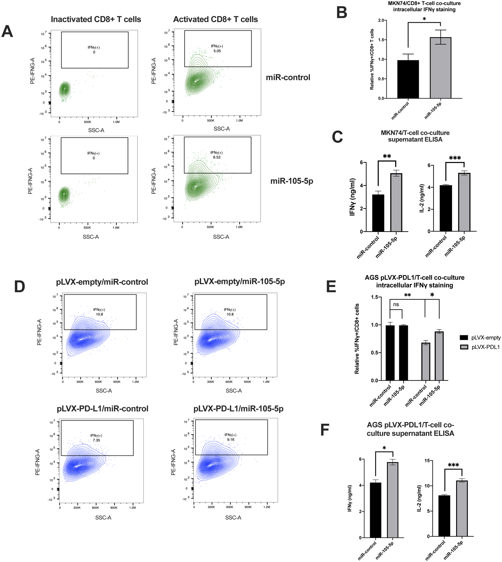
Figure 5. Overexpression of miR-105-5p in cancer cells downregulates PD-L1 and promotes increased activation of CD8+ T cells in an in vitro co-culture system.

(A) Representative flow cytometry plots showing the percentage of IFNγ-positive CD8+ T cells in a co-culture experiment with miR-control or miR-105-5p transfected MKN74 cells. The cells were pre-selected for being CD3+CD8+. (B) Graph represents the relative percentage of IFNγ-positive CD8+ T cells in miR-control vs miR-105-5p transfected MKN74 cells in three independent experiments. (C) Supernatant collected from MKN74/CD8+ T cell co-culture experiments was analyzed with ELISA assays for the levels of secreted IFNγ and IL-2. (D) Representative flow cytometry plots of the IFNγ-positive CD8+ T cells in a co-culture experiment with pLVX-control or pLVX-PD-L1 transduced AGS cells transfected with miR-control or miR-105-5p transfected. (E) Graph represents the mean of three independent experiments described in (D). (F) Supernatant from co-culture experiments described in (D) were analyzed by ELISA for the levels of secreted IFNγ and IL-2 from activated CD8+ T cells. All experiments were repeated in triplicate and bar plots represent the mean ± SD of all experiments. Significance was calculated by Student’s t-test, *P < 0.05; **P < 0.01; ***P < 0.001.
