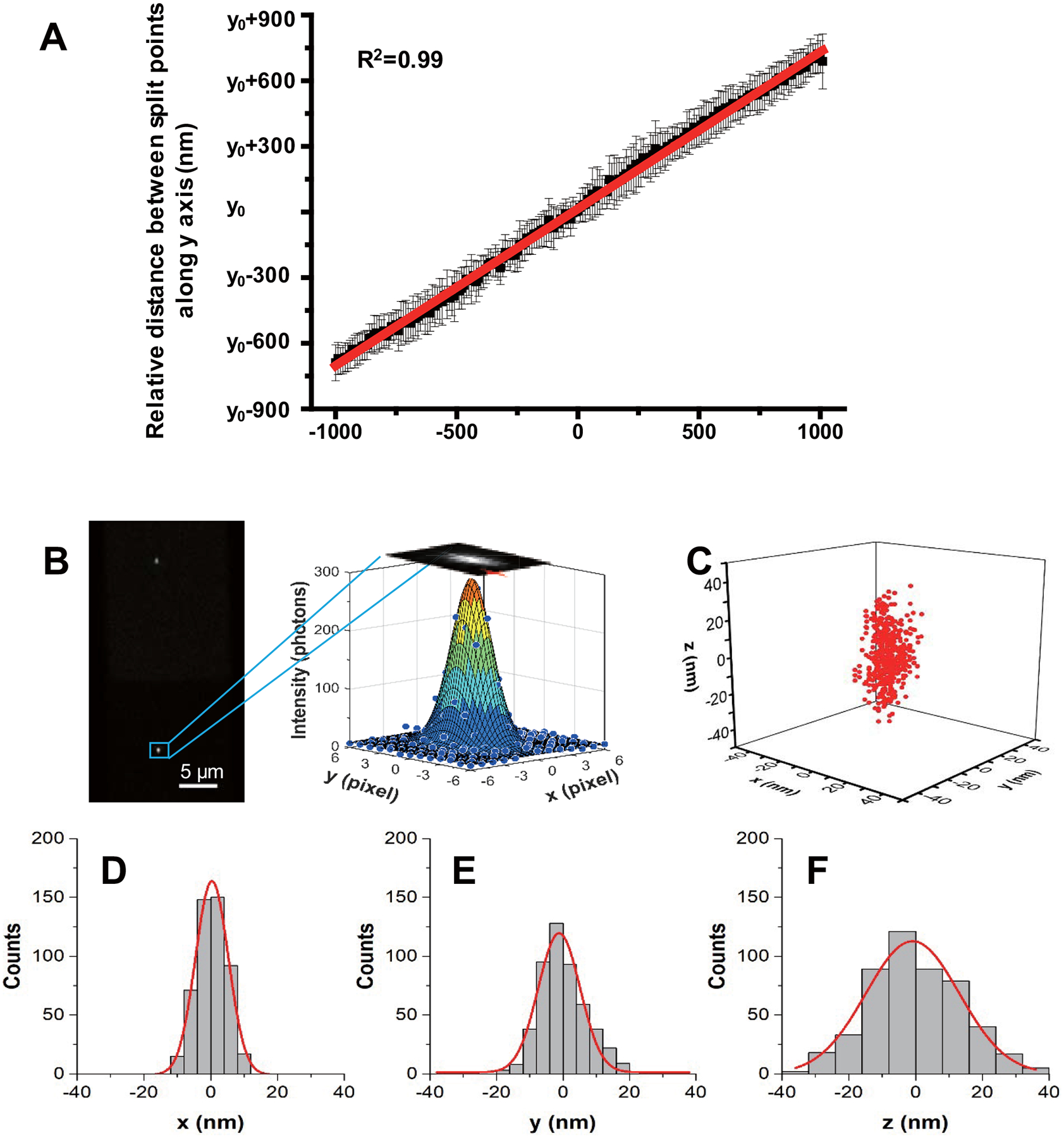
Extended Data Fig. 1. Calibration curve of Δy vs. Δz and 3D localization precision of a AuNR.

(A) The AuNRs were immobilized on a glass slide surface with various orientations and scanned along the z-axis from −1000 nm to 1000 nm with 10 nm steps using a high-precision objective scanner (Data were expressed as mean ± SD, n=20 independent experiments). (B) Typical upper and lower half-plane dark-field images of a AuNR with 0.02 s integration time are shown on the left. Scale bar is 5 μm. The lateral positions of the AuNR are determined by 2D elliptical Gaussian fitting (right) the intensity profile. (C) Scatter plot of locations of the same AuNR in 500 frames. The x, y positions are determined using 2D elliptical Gaussian fitting of the particle image intensity profile. The z positions are obtained from feedback of the objective scanner when auto-focusing system was engaged. The localization precision is determined as the standard deviation from 1D Gaussian function fitting the histogram distribution of the AuNR locations in x, y, z, giving σx = 4.9 nm (D), σy = 6.3 nm (E) and σz = 14.0 nm (F).
