Fig. 1.

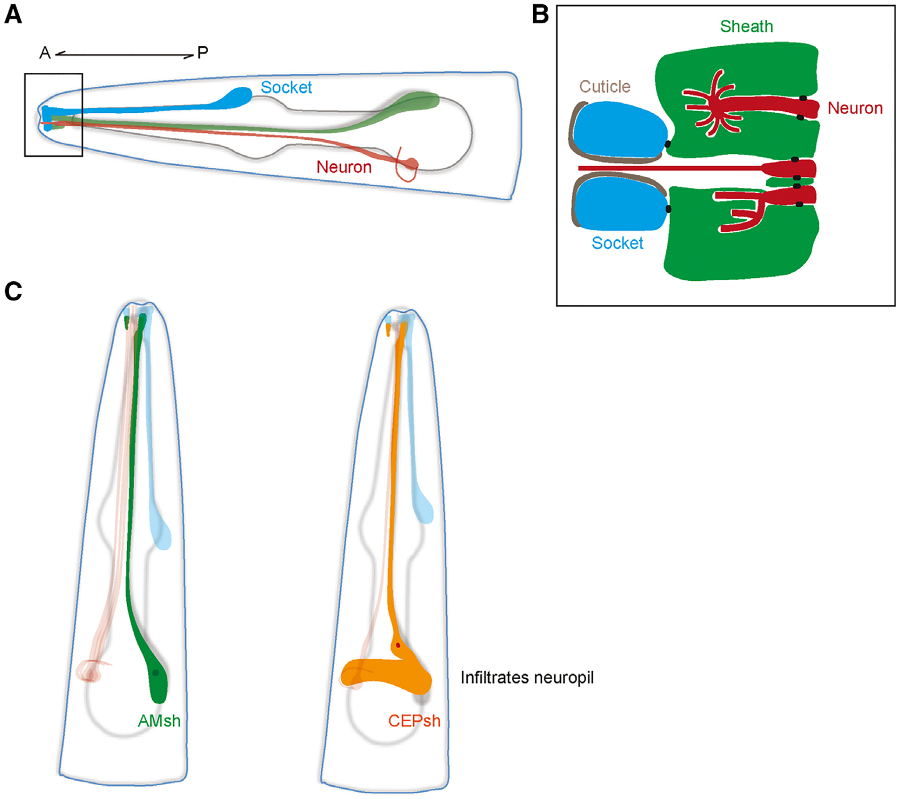
(A) Schematic of a generic sense organ in C. elegans. The processes of socket (blue) and sheath glia (green) together form a channel at the nose tip where dendrites of sensory neurons (red) protrude through to interact with the environment. A, anterior; P, posterior. (B) Close-up of the channel at the nose tip area demarcated by the black box in A. Sheath glia (green) are connected to sensory neurons (red) and socket cells (blue) by apical junctions (black dots). Socket cells are connected to the cuticle of the worm. The cilia may vary in shape depending on the type of neuron. The dendrites of some neurons remain in the channel while others can be completely or partially embedded in the sheath glia. (C) Schematic of AMsh (left) and CEPsh (right) glia. Note that the posterior process of the CEPsh infiltrates the neuropil and comes into close contact with synapses like astrocytes do.
